

Foot Muscles
The human foot is essential for walking, running, and all other forms of locomotion. There are about twenty intrinsic muscles and several extrinsic muscles that work together to move and stabilize the foot. They control toe movement, maintain the arches, absorb shock, and provide balance and propulsion.
The foot’s stability also depends on the plantar fascia, a strong band of connective tissue linking the bones, muscles, and soft tissues on the sole. Working with tendons and ligaments, it supports the arches and keeps the foot firm during standing and movement.
Intrinsic Muscles of the Foot
The intrinsic muscles both originate and insert within the foot and are often the primary group considered when describing the muscles of the foot. They are responsible for fine movements of the toes, maintaining the arches of the foot, and providing stability and balance during standing and walking.
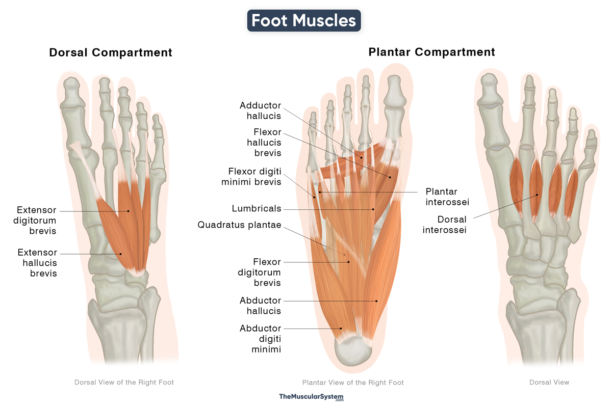
Intrinsic Foot Muscles
They are organized into two main compartments — dorsal and plantar. The plantar compartment is further divided into four layers. Below is a list of all the intrinsic muscles of the foot, along with their location and action.
| Muscle | Origin | Insertion | Action | Innervation | Blood Supply |
|---|---|---|---|---|---|
| Dorsal Compartment: Contains muscles located on the dorsum or top of the foot. | |||||
| Extensor hallucis brevis | Dorsal surface of the calcaneus bone | Base of the proximal phalanx of the big toe | Extends the 1st, or big toe | Deep fibular nerve (L5-S1) | Dorsalis pedis artery |
| Extensor digitorum brevis | Upper and lateral surfaces of the calcaneus bone | Lateral sides of the 2nd-4th extensor digitorum longus tendons | Extends the 2nd-4th toes | Deep fibular nerve (L5-S1) | Dorsalis pedis, and fibular arteries |
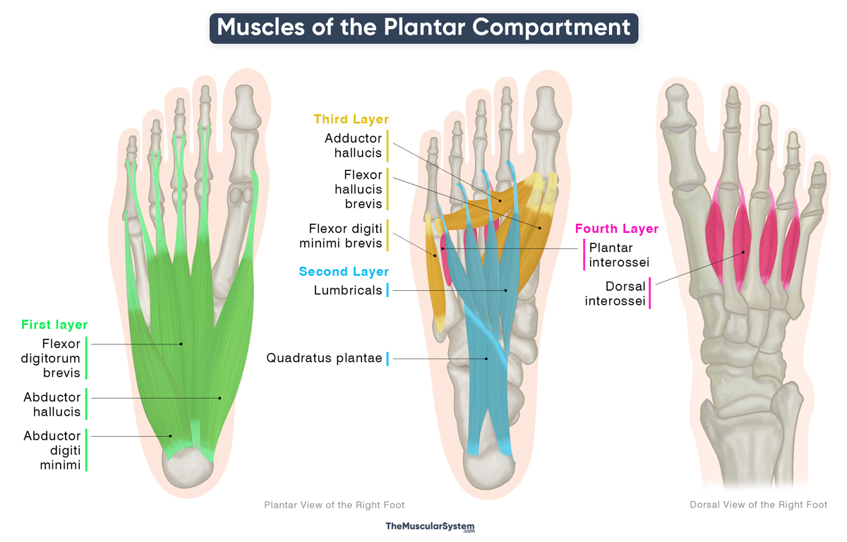
| Plantar Compartment: Contains muscles located on the sole or bottom of the foot | |||||
| First layer | |||||
| Abductor hallucis | Medial process of calcaneal tuberosity, flexor retinaculum, and plantar aponeurosis | Medial side of the base of the first proximal phalanx (of the big toe) | — Abducts and flexes the big toe— Supports the medial longitudinal arch | Medial plantar nerve (S1-S3) | Medial plantar, and first plantar metatarsal artery |
| Flexor digitorum brevis | Medial process of the calcaneal tuberosity, the plantar aponeurosis, and the intermuscular septum of the foot | Medial and lateral surfaces of the 2nd to 5th middle phalanges | — Flexes the 2nd-5th toes— Supports the longitudinal arch of the foot | Medial plantar nerve (S1-S3) | Medial plantar, lateral plantar, plantar metatarsal, and common plantar digital arteries |
| Abductor digiti minimi of the foot | Lateral and medial processes of the calcaneal tuberosity, and the plantar aponeurosis | Proximal phalangeal base of the 5th or little toe | Abducts and flexes the 5th, or little toe | Lateral plantar nerve (S1-S3) | Lateral plantar artery |
| Second layer | |||||
| Quadratus plantae | — Medial head: Medial side of the plantar surface of the calcaneus bone— Lateral head: Lateral side of the plantar surface of the calcaneus bone | The flexor digitorum longus tendon | Assists the flexor digitorum longus in flexing the 2nd -5th toes | Lateral plantar nerve (S1-S3) | Medial, and lateral plantar arteries |
| Lumbricals of the foot (Group of 4 muscles) | Medial and lateral sides of the 1st to 4th flexor digitorum longus tendons | The 2nd to 5th proximal phalangeal base and the attached extensor hoods | Flexes and extends the 2nd-5th toes at different joints | 1st lumbrical: Medial plantar nerve (S2-S3)2nd-4th lumbricals: Lateral plantar nerve (S2-S3) | Medial, and lateral plantar arteries |
| Third Layer | |||||
| Flexor hallucis brevis | — Lateral head: Medial plantar surface of the cuboid, and the adjacent lateral cuneiform— Medial head: Plantar part of the tibialis posterior tendon | Lateral and medial sides of the proximal phalangeal base of the big toe | — Flexes the big toe— Supports the medial longitudinal arch | Medial plantar nerve (S1-S2) | Medial plantar, and first metatarsal artery |
| Adductor hallucis | — Oblique head: The 2nd-4th metatarsal base— Transverse head: Plantar metatarsophalangeal ligaments of the 3rd-5th toes | Lateral side of the proximal phalangeal base of the big toe | — Adducts the big toe— Supports the transverse arch | Lateral plantar nerve (S2-S3) | Medial, and lateral plantar arteries |
| Flexor digiti minimi brevis of the foot | Medial side of the plantar surface of the 5th metatarsal’s base | Lateral side of the fifth proximal phalangeal base | — Flexes the 5th toe— Supports the lateral longitudinal arch | Lateral plantar nerve (S2-S3) | Lateral plantar, arcuate, and lateral tarsal arteries |
| Fourth Layer | |||||
| Plantar interossei (Group of 3 muscles) | Medial-plantar surface of the 3rd to 5th metatarsal | Medial side of the proximal phalangeal base of the 3rd-5th toes | Flexes, abducts, and extends the 3rd-5th toes at different joints | Lateral plantar nerve (S2-S3) | Lateral plantar artery |
| Dorsal interossei (Group of 4 muscles) | Sides of the proximal half of the 1st-5th metatarsal bones | Bases of the 2nd-4th proximal phalanges of the lateral toes | Flexes, abducts, and extends the 2nd-4th toes at different joints | Lateral plantar nerve (S2-S3) | Dorsalis pedis, dorsal metatarsal, plantar metatarsal, and lateral plantar arteries |
Plantar Muscles at the Bottom of Foot
Apart from the above vertical classification, the plantar compartment can also be divided horizontally, from the medial to the lateral sides.
Medial plantar muscles: These are the muscles that insert into the hallux or big toe, and mainly act to move this toe.
- Abductor hallucis
- Adductor hallucis
- Flexor hallucis brevis
Lateral plantar muscles: These are the muscles that insert into the 5th digit or little toe, and mainly work to move this toe.
- Abductor digiti minimi
- Flexor digiti minimi
Central plantar muscles: These are the muscles that insert into the second to fourth or fifth digits, and act on these same digits.
- Lumbricals
- Quadratus plantae
- Flexor digitorum brevis
- Dorsal interossei
- Plantar interossei
Extrinsic Muscles of the Foot
The extrinsic foot muscles originate in the lower leg (tibia, fibula, or femur) and insert into the bones of the foot. They cross the ankle joint to produce the major movements of the foot: dorsiflexion, which lifts the foot upward; plantarflexion, which points it downward; inversion, which turns the sole inward; and eversion, which turns it outward. These muscles provide power and stability during walking and running, working together with the intrinsic muscles to maintain balance and control.
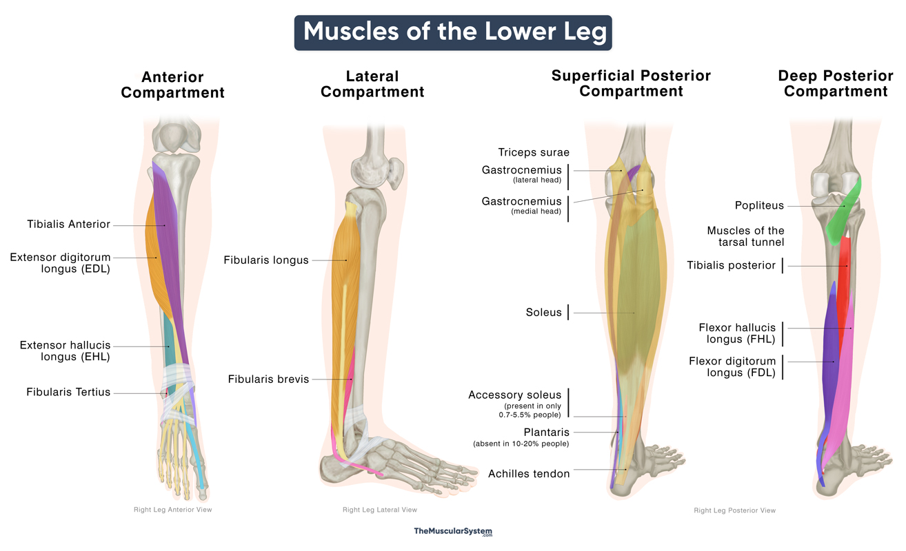
Lower Leg Muscles
They are anatomically classified as lower leg muscles and are divided into four groups based on where they are located. Here is a list of the extrinsic muscles of the foot, and their action:
| Muscles | Actions |
|---|---|
| Anterior Compartment: Contains muscles located at the front of the lower leg | |
| Tibialis anterior | Dorsiflexes and inverts the foot |
| Extensor hallucis longus | — Extends the big toe— Dorsiflexes the foot |
| Extensor digitorum longus | — Extends the lateral four toes— Dorsiflexes the foot |
| Fibularis tertius | Dorsiflexes and everts the foot |
| Lateral Compartment: Contains muscles located on the outer or fibular side of the lower leg | |
| Fibularis longus | — Everts and weakly plantarflexes the foot— Supports the transverse arch |
| Fibularis brevis | — Everts and weakly plantarflexes the foot— Supports the medial longitudinal arch |
| Posterior Compartment (Superficial Group): Contains muscles located at the back of the lower leg, forming the fleshy calf region just below the knee joint | |
| Gastrocnemius | — Plantarflexes the foot— Flexes the knee |
| Soleus | Plantarflexes the foot |
| Plantaris | — Weakly plantarflexes the foot— Flexes the knee |
| Posterior Compartment (Deep Group): Contains muscles lying deep at the back of the lower leg | |
| Tibialis posterior | — Plantarflexes and inverts the foot— Supports the medial arch |
| Flexor hallucis longus | — Flexes the big toe— Plantarflexes the foot— Supports the medial longitudinal arch |
| Flexor digitorum longus | — Flexes the lateral four toes— Plantarflexes the foot |
Note: The popliteus, in the deep posterior compartment, extends from the knee to the tibia and acts only on the knee. It is the only lower leg muscle that does not act on the foot.
References
- Anatomy, Bony Pelvis and Lower Limb, Foot Muscles: NCBI.NLM.NIH.gov
- Muscles of the Foot: TeachMeAnatomy.info
- Ankle and Foot Anatomy: Kenhub.com
- Muscles of the Lower Leg and Foot: Bio.Libretexts.org
- Anatomy of the Foot: Arthritis.org


