

Hand Muscles
Hands are the most distal part of the upper limbs – but their functioning involves almost everything humans do, from everyday tasks like eating, writing, and driving to playing baseball, fishing, and rock climbing.
The opposable thumbs in human hands make it possible to perform all these tasks. Several muscles in the hands help flex and extend the thumb and fingers. They also help with gripping things and making a fist – basically, anything you do with your hands. These muscles can be divided into two groups based on their point of origin – intrinsic and extrinsic.

Hand Muscle
There are 11 intrinsic muscles and 15 extrinsic muscles in each hand.
Names of the Muscles of the Hand and Fingers With Basic Anatomy
Intrinsic Muscles in the Hand
These muscles originate from the hand’s bones, ligaments, or fascia, also getting inserted within the hand area. It means these are the muscles that are located in their entirety in the hand region.
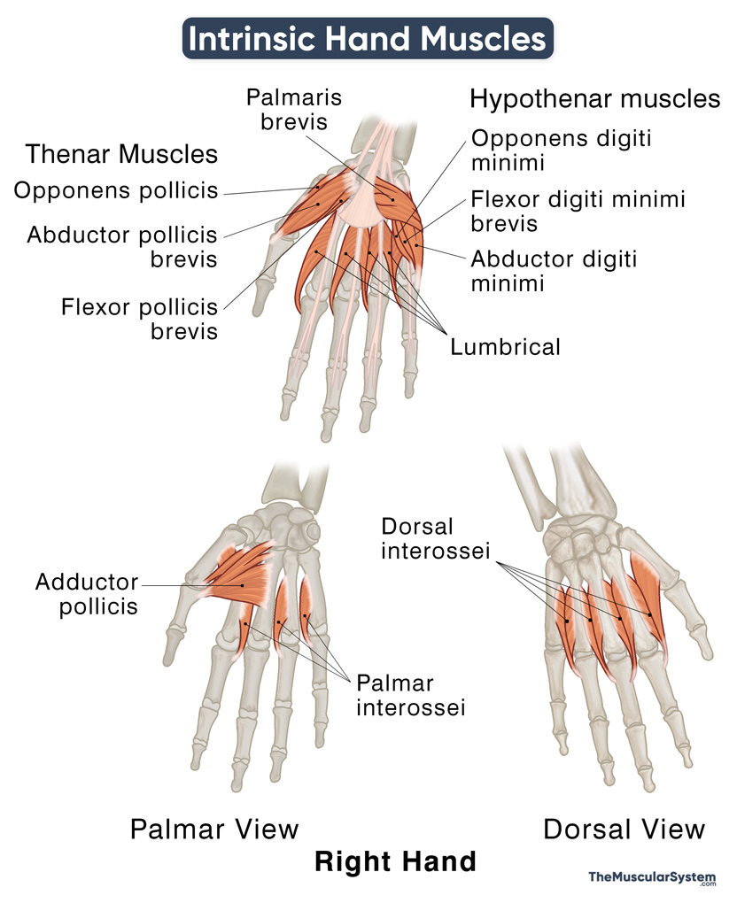
Intrinsic Muscles of the Hand
These are primarily involved in further divided into multiple groups based on their location:
Thenar Muscles
These are the muscles that form the thenar eminence of the palm. There are 3 muscles in this group, all located in the thenar region at the base of the thumb. These muscles help with most of the significant thumb movements.
| Muscle | Origin | Insertion | Action | Innervation |
| 1. Opponens pollicis | Tubercle of the trapezium and adjacent flexor retinaculum | Radial surface of the 1st metacarpal | Flexion, adduction, and rotation of the thumb at its carpometacarpal joint (thumb opposition) | Recurrent branch of median nerve (C8, T1) |
| 2. Flexor pollicis brevis | Superficial head: Tubercle of the trapezium and adjacent flexor retinaculum
Deep head: Trapezoid and capitate |
Radial surface of the 1st proximal phalangeal base | Thumb flexion at its metacarpophalangeal joint | Recurrent branch of median nerve (C8, T1); Deep branch of ulnar nerve (deep head) |
| 3. Abductor pollicis brevis | Flexor retinaculum and the tubercles of the scaphoid and trapezium | Radial surface of the 1st proximal phalangeal base | Abduction of the thumb at its carpometacarpal and metacarpophalangeal joints | Recurrent branch of median nerve (C8, T1) |
Hypothenar Muscles
The hypothenar muscles form the hypothenar eminence, or the fleshy mound on the palm’s ulnar side, at the little finger’s base. Like the thenar muscles, there are 3 hypothenar muscles in each hand. Their primary function is to help with the movements of the little finger.
| Muscle | Origin | Insertion | Action | Innervation |
| 4. Abductor digiti minimi | Pisiform bone, pisohamate ligament, and flexor retinaculum | Ulnar surface of the 5th proximal phalangeal base | Abduction of the little finger at its metacarpophalangeal joint | Deep branch of ulnar nerve (C8, T1) |
| 5. Flexor digiti minimi brevis | Hook of hamate and adjacent flexor retinaculum | Ulnar surface of the 5th proximal phalangeal base | Flexion of the little finger at its metacarpophalangeal joint | Deep branch of ulnar nerve (C8, T1) |
| 6. Opponens digiti minimi | Hook of hamate and adjacent flexor retinaculum | Ulnar surface of the 5th proximal phalanx | Rotation and opposition of the little finger at its Carpometacarpal joint | Deep branch of ulnar nerve (C8, T1) |
The lumbricals are a group of 4 narrow muscles in the metacarpal area of the hand. These are attached to the 2nd to 5th digits of the hand, helping with finger flexion and extension.
| Muscle | Origin | Insertion | Action | Innervation |
| 7. Lumbricals | 1st: Flexor digitorum profundus tendon of the 2nd digit
2nd: FDP tendon of the 3rd digit 3rd: FDP tendon of the 3rd and 4th digits 4th: FDP tendon of the 4th and 5th digits |
The radial margin of dorsal digital expansion of the extensor digitorum at the 2nd to 5th proximal phalanx (respectively) | Finger flexion at 2nd to 5th metacarpophalangeal joints; Finger extension at the 2nd to 5th interphalangeal joints | 1st and 2nd: Digital branch of median nerve (C8-T1)
3rd and 4th: Deep branch of ulnar nerve (C8-T1) |
Interossei
The interossei are small, worm-like muscles located between the metacarpal bones. They are divided into two groups based on their location – dorsal and palmar Interossei. Each hand has 4 dorsal and 3 (sometimes 4) palmar interossei. They help with abduction, adduction, flexion, and extension of the fingers.
| Muscle | Origin | Insertion | Action | Innervation |
| 8. Dorsal interossei | 1st: Palmar and ulnar side of 1st metacarpal’s base; Radial border of the 2nd or the index finger’s metacarpal
2nd: Ulnar border of 2nd metacarpal; Radial border of 3rd metacarpal 3rd: Ulnar border of 3rd metacarpal; Radial border of 4th metacarpal 4th: Ulnar border of 4th metacarpal; Radial border of 5th metacarpal |
2nd to 4th digit’s proximal phalangeal base and adjacent extensor | Finger abduction and flexion at the metacarpophalangeal joint; finger extension at the interphalangeal joints | Deep branch of ulnar nerve (C8 and T1) |
| 9. Palmar interossei | 1st (not always present): Ulnar surface of 1st metacarpal’s base
2nd: Ulnar surface of 2nd metacarpal 3rd: Radial surface of 4th metacarpal 4th: Radial surface of 5th metacarpal |
1st to 4th proximal phalangeal base and adjacent extensor expansion | Finger adduction and flexion at 2nd, 3rd and 5th metacarpophalangeal joints; Finger extension at 2nd, 3rd and 5th interphalangeal joints | Deep branch of ulnar nerve (C8 and T1) |
Other Intrinsic Hand Muscles
There are 2 more muscles in the hand that do not belong to any of the above groups, though they do work with the other hand muscles to help with the functioning of the hands.
| Muscle | Origin | Insertion | Action | Innervation |
| 10. Adductor pollicis | Oblique head: 2nd and 3rd metacarpal bases and the capitate bone
Transverse head: Palmar side of 3rd metacarpal’s shaft |
Medial side of 1st proximal phalangeal base, | Thumb adduction at its carpometacarpal joint | Deep branch of the ulnar nerve (C8 and T1) |
| 11. Palmaris brevis | Flexor retinaculum and palmar aponeurosis | Dermis of the skin in the hypothenar region | Strengthening the grip of hand by tightening the palmar aponeurosis | Superficial branch of the ulnar nerve (C8, T1) |
Mnemonics
Mnemonic 1
Here is a mnemonic that helps remember all the thenar and hypothenar muscles along with the adductor pollicis, which is located in the thenar region. Here are the muscles, from the radial to the ulnar side:
All For One And One For All
- Abductor pollicis brevis
- Flexor pollicis brevis
- Opponens pollicis
- Adductor pollicis
- Opponens digiti minimi
- Flexor digiti minimi brevis
- Abductor digiti minimi
The palmaris brevis (located in the hypothenar region) is the only muscle not included.
Mnemonic 2
This one helps remember the intrinsic muscles that receive innervation from the median nerve.
FOAL
- Flexor pollicis brevis
- Opponens pollicis
- Abductor pollicis brevis
- Lateral two lumbricals
The rest of the intrinsic muscles are innervated by the ulnar nerve.
Extrinsic Muscles in the Hand
These muscles do not originate within the hand, but they have their points of insertion in the bones or fascia of the hand. As a result, they are vital to the functioning of the hand and are some of the primary muscles that help move the fingers and thumb.
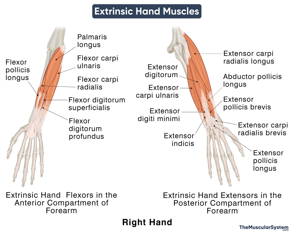
Extrinsic Muscles of the Hand
All the extrinsic muscles of the hand originate in the forearm and then course distally to enter the hand. These can be divided into two groups based on their action: the flexors and extensors.
Extrinsic Flexors of the Hand in the Anterior Compartment of the Forearm
Superficial Flexors
All three superficial flexors insert in the carpal and metacarpal areas of the hand and help with flexion, as well as radial and ulnar deviation of the wrist.
Intermediate Flexors
Deep Flexors
- Flexor digitorum profundus
- Flexor pollicis longus
The intermediate and deep flexors all insert into the phalangeal region of the hand. The actions of these muscles include flexing the wrist and the fingers at the metacarpophalangeal and interphalangeal joints.
Extrinsic Extensors of the Hand in the Posterior Compartment of the Forearm
Superficial Extensors
- Extensor digitorum
- Extensor digiti minimi
- Extensor carpi ulnaris
- Extensor carpi radialis longus
- Extensor carpi radialis brevis
These muscles insert into the dorsal side of the hand – into the metacarpal bones or in the extensor expansion. Their actions include extending the wrist and the fingers at their metacarpophalangeal and interphalangeal joints. The extensor carpi ulnaris adducts the wrist, while the extensor carpi radialis longus and brevis both help with wrist abduction.
Deep Extensors
These muscles insert at different points into the 1st and 2nd digits. The extensor indicis helps with the extension of the index finger, while the rest of the muscles in this group help with the extension and abduction of the thumb at its different joints.
References
- Anatomy, Shoulder and Upper Limb, Hand Muscles: NCBI.NLM.NIH.gov
- Hand Muscles: ASSH.org
- Muscles of the Hand: Osmosis.org
- Muscles of the Hand: TeachMeAnatomy.info
- Hand Anatomy: KenHub.com
- Anatomy, Shoulder and Upper Limb, Hand Intrinsic Muscles: StatPearls.com
- Intrinsic Muscles of the Hand: WheelessOnline.com
- Extrinsic muscles of the hand: RadioPaedia.org


