

Flexor Digitorum Profundus
By
Della Barnes, an MS Anatomy graduate, blends medical research with accessible writing, simplifying complex anatomy for a better understanding and appreciation of human anatomy.
Last updated:
05/05/2023Della Barnes, MS Anatomy
UX/UI Designer at - AdobeDella Barnes, an MS Anatomy graduate, blends medical research with accessible writing, simplifying complex anatomy for a better understanding and appreciation of human anatomy.
What is the Flexor Digitorum Profundus
The flexor digitorum profundus, or FDP, is a muscle in the deep anterior compartment of the forearm. It is one of the muscles forming the third muscular layer in the anterior compartment. The other one is the deep muscle, flexor pollicis longus.
As the name implies, the flexor digitorum profundus is involved in finger flexion. It is one of the carpal tunnel tendons.
Anatomy
Location and Attachments
| Origin | Proximal 3/4 of the ulna, from the anterior and medial surfaces; the adjacent part of the interosseous membrane |
| Insertion | The palmar surface of the distal phalangeal bases of 2nd to 5th digits |
The flexor digitorum profundus has an origin that spreads over a broad region. The primary point of origin is the front and medial surfaces of the top (proximal) 3/4 of the ulna, on the side of the small finger, via the common extensor tendon. It shares this origin point with the extensor and flexor carpi ulnaris muscles. The origin also includes the olecranon and coronoid processes of the ulna. Some fibers rise from the adjoining interosseous membrane and the forearm’s deep fascia.
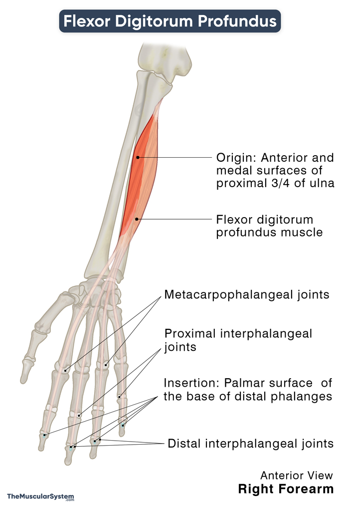
After originating, the tendons widen and form the belly of this fusiform muscle, coursing downward or distally towards the hand and fingers. The muscle gives rise to a broad tendon at the distal end of the ulna, near the wrist joint. It then travels superficial to the pronator quadratus and deep to the flexor retinaculum, the fibrous band in the palm.
Once it reaches the palm, the thick tendon branches into 4 slips. Together they pass through the carpal tunnel, with the tendon for the index finger (2nd digit) separating from the other 3 partially attached tendons around this point. Afterward, the tendons insert into the palmar or volar surface of the base of the distal phalanges of the index, middle, ring, and small fingers (2nd to 5th digits).
Relations With Surrounding Muscles and Structures
As mentioned above, it is one of the three deep anterior forearm muscles, along with the flexor pollicis longus and pronator quadratus. Within the deep compartment, the flexor digitorum profundus and flexor pollicis longus form the upper layer, with the pronator quadratus forming the deepest layer.
The flexor digitorum profundus lies deep to the anterior muscle, flexor digitorum superficialis, throughout its course. The 4 insertion tendons of this muscle decussate the flexor digitorum superficialis at the point where the latter’s superficial and deep tendons slip together (Camper chiasm) before passing into the carpal tunnel.
Functions
| Action | Flexion of the distal interphalangeal (DIP) joints of the 2nd-5th digits; flexion of the proximal interphalangeal (PIP), metacarpophalangeal (MCP), and wrist joints (secondary) |
Flexing the Fingers
This muscle is one of the main flexors of the fingers. The deep surface of each flexor digitorum profundus tendon acts as the point of attachment for the corresponding lumbrical muscle of the hand, which then insert into the extensor hood of the proximal phalanges. So, the FDP and the lumbricals work in synergy to flex the 2nd to 5th fingers at their distal interphalangeal joints.
The other synergists of FDP in flexing the fingers are the flexor digitorum superficialis and flexor digiti minimi brevis.
It also plays a secondary role in flexing these fingers at their proximal interphalangeal and metacarpophalangeal joints.
Since it helps flex the 2nd to 5th fingers, this muscle plays a significant role in enabling us to grip something, and maintain the strength of the grip.
Flexing the Wrist
The muscle helps with the functioning of the primary wrist flexors, the flexor carpi radialis, flexor carpi ulnaris, palmaris longus, flexor pollicis longus, and flexor digitorum superficialis. This is why the FDP is considered an accessory flexor of the wrist.
The primary antagonist of the flexor digitorum profundus is the superficial posterior compartment muscle extensor digitorum.
Innervation
| Nerve | Median nerve, via anterior interosseous branch (C8 and T1), for the lateral part Ulnar nerve (C8 and T1) for the medial part |
The muscle has dual innervation, one each for its lateral and medial parts. Its lateral part refers to the portion of the muscle that attaches to the index and middle fingers; it is innervated by the anterior interosseous branch (C8, T1) of the median nerve.
The medial part is the portion that attaches to the ring and little fingers. The ulnar nerve (C8, T1) innervates this part of the muscle.
The middle finger often receives nerve supply from both the above sources.
Blood Supply
| Artery | Ulnar artery branches |
The inferior ulnar collateral artery and the ulnar recurrent artery supply the muscle at its point of origin. The upper part of the muscle belly gets its vasculature from the common interosseous artery, a branch of the ulnar artery. The rest of the muscle receives its arterial supply from the anterior interosseous artery, as well as other branches of the ulnar and medial arteries.
References
- Flexor Digitorum Profundus: Origin, Action & Insertion: Study.com
- Flexor Digitorum Profundus: TeachMeAnatomy.info
- Flexor Digitorum Profundus Muscle: Radiopaedia.org
- Flexor Digitorum Profundus Muscle: KenHub.com
- Flexor Digitorum Profundus: Rad.Washington.edu
- Body Anatomy: Upper Extremity Tendons: ASSH.org
Della Barnes, an MS Anatomy graduate, blends medical research with accessible writing, simplifying complex anatomy for a better understanding and appreciation of human anatomy.
- Latest Posts by Della Barnes, MS Anatomy
-
Tensor Tympani
- -
Stapedius
- -
Auricularis Posterior
- All Posts


