

Abdominal Muscles
The abdominal muscles are a group of paired skeletal muscles that cover the front and sides of the abdomen, positioned symmetrically along the spine. These muscles are vital in protecting internal organs, providing structural integrity, supporting posture, and helping with torso movements.
In this context, “abdominal muscles” refers specifically to the skeletal muscles that construct the outer abdominal wall and are not to be confused with the smooth muscles of internal organs such as those in the digestive tract. Together with surrounding connective tissues and fascia, they form the structural core of the abdominal wall.
Structure of the Abdominal Wall
The human abdominal wall is composed of multiple layers, including the skin, fascia, muscles, and connective tissues, which together enclose the abdominal cavity. It is the largest cavity in the human body, extending from the ribs and diaphragm at the top to the pelvis at the bottom.
From superficial to deep, the layers of the abdominal wall are:
- Skin
- Superficial fascia
- Camper’s fascia (fatty layer)
- Scarpa’s fascia (membranous layer)
- Abdominal muscles (of the anterolateral abdominal wall)
- Transversalis fascia
- Extraperitoneal fat
- Parietal peritoneum (outer layer of the peritoneum, the serous membrane lining the abdominal cavity)
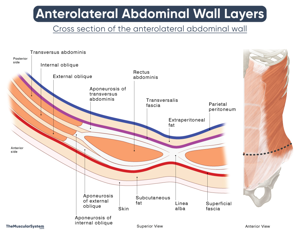
Layers of the Anterolateral Abdominal Wall
Unlike the thoracic and pelvic regions, the abdominal wall lacks extensive bony support, except for the lower ribs above and the lumbar vertebrae in the back. This relative absence of rigid structures allows the abdominal wall to serve as a flexible connection between the thoracic cage and pelvis. It also enhances trunk mobility, a function supported by the muscles in the region. The abdominal muscles attach to the ribs, vertebral column, and pelvic bones, playing key roles in trunk and hip movement.
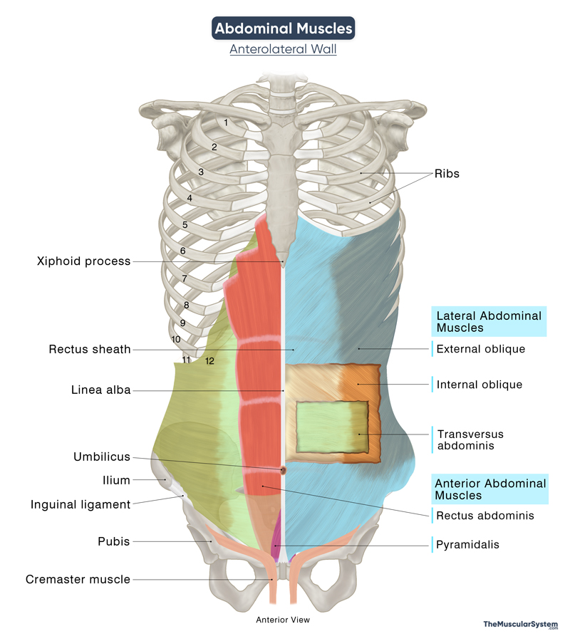
Name, Location, and Anatomy of the Muscles of the Abdomen
They are typically divided into two groups: anterolateral abdominal wall muscles and posterior abdominal wall muscles. Some sources classify only the five anterolateral muscles as abdominal muscles, placing the posterior group within the category of pelvic muscles due to their involvement in spinal and pelvic stability and movements.
Here, all five anterolateral and four posterior muscles are considered in the group of abdominal muscles. The anterolateral group is further subdivided into anterior and lateral abdominal wall muscles, based on their anatomical positioning.
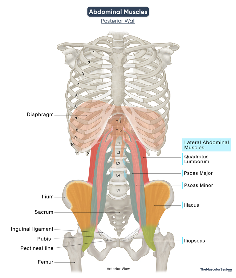
Abdominal Muscles of Posterior Wall
Find out more about the anatomy and function of each muscle:
| Name | Location | Origin | Insertion | Action | Innervation | Blood Supply |
|---|---|---|---|---|---|---|
| Muscles of the Anterolateral Abdominal Wall | ||||||
| Anterior Abdominal Wall | ||||||
| Rectus Abdominis | The most superficial muscle; it runs vertically down the midline from ribs to pubic bone, under the belly button | Pubic crest, tubercle, and symphysis | Xiphoid process, and the 5th to 7th costal cartilages | – Trunk flexion
– Abdominal compression to help with bodily functions like urination, labor – Core stability |
T7 to T11 spinal nerves, subcostal nerve (T12) | Superior and Inferior epigastric arteries |
| Pyramidalis | Triangular muscle that sits on the pubic bone in the lower midline of the abdomen | Pubic crest and symphysis | Linea alba | – Helps tighten the linea alba to maintain abdominal wall integrity | Subcostal nerve (T12) | Inferior epigastric artery |
| Lateral Abdominal Wall | ||||||
| External Oblique | Covers the sides of the abdomen from lower ribs to iliac crest, slanting downward | 5th-12th ribs’ safts | Iliac crest, pubic tubercle, ASIS, and linea alba | – Trunk flexion and rotation
– Core stability |
Thoraco abdominal (T7-T11), and subcostal (T12) nerves | Lower posterior intercostal, subcostal, and deep circumflex iliac arteries |
| Internal Oblique | Sits beneath external oblique, slanting upward from iliac crest to lower ribs | Inguinal ligament, iliac crest, thoracolumbar fascia | 10th-12th ribs’ lower borders, linea alba, pubic crest, pectineal line | – Trunk flexion and rotation
– Abdominal compression to help with bodily functions |
Thoracoabdominal (T7-T11), subcostal (T12), ilioinguinal (L1), and iliohypogastric (L1) nerves | Posterior intercostal, and subcostal arteries |
| Transversus Abdominis | Deepest abdominal muscle, runs horizontally from the spine to the midline of the abdomen | Inguinal ligament, iliac crest, thoracolumbar fascia, 5th-10th ribs’ costal cartilage | Linea alba, pubic crest, pectineal line of the pubis | – Abdominal compression to help with bodily functions
– Core stability |
Thoracoabdominal (T7-T11), subcostal (T12), ilioinguinal (L1), and iliohypogastric (L1) nerves | Lower posterior intercostal and subcostal arteries |
| Muscles of the Posterior Abdominal Wall | ||||||
| Quadratus Lumborum | Deep in the posterior abdominal wall, between the 12th rib and the iliac crest | Iliac crest’s border, iliolumbar ligament | L1-L4 vertebral transverse processes, 12th rib’ lower border | – Lateral trunk flexion
– Lower back extension and stability – 12th rib stabilization to aid breathing |
Subcostal (T12) and L1-L4 lumbar nerves | Subcostal, iliolumbar, lumbar, and median sacral arteries |
| Iliopsoas
Formed by the blending of the following two muscles: |
||||||
| — Psoas Major | Runs from lumbar spine through pelvis to upper thigh, near the inguinal region | T12-L4 vertebral bodies, transverse processes, intervertebral disks | Lesser trochanter of the femur | Hip flexion and rotation | L1-L3 spinal nerves | Lumbar branch of iliolumbar artery |
| — Iliacus | Fills the iliac fossa, running from the pelvis to the lumbar spine | Iliac fossa, iliac crest | Lesser trochanter of the femur | Hip flexion | Femoral nerve | Lumbar branch of iliolumbar artery |
| Psoas Minor | In front of the psoas major in the lower lumbar region (often absent) | T12 and L1 vertebral bodies, intervertebral disks | Iliopubic eminence, pectineal line | Flexion of the lumbar region | L1 spinal nerve | Lumbar arteries |
The muscles of the lateral abdominal wall (external oblique, internal oblique, transversus abdominis)
The internal oblique muscle extends downward into the pelvic region and gives rise to the cremaster muscle, which is fully developed only in males and helps raise the testes. Since it doesn’t directly contribute to the abdominal wall, it’s not included in the table above.
FAQ
Do women have different abdominal muscles from men?
The abdominal muscles are the same in both men and women, but differences in hormones, fat distribution, and pelvic structure can influence how they look. For example, in women, higher estrogen levels promote the storage of subcutaneous fat, the type of fat located directly beneath the skin. Additionally, women may experience changes in the structure of their abdominal muscles during pregnancy (e.g., diastasis recti). In contrast, men often develop more defined abdominal musculature due to lower body fat and higher testosterone levels.


