

Perineal Muscles
What is the Perineum
In general terms, the perineum is the area of skin between the genitals and the anus. In anatomy, it refers to a deeper region that includes muscles, nerves, and vessels of the urogenital and anal areas located at the bottom of the pelvis, just beneath the pelvic floor.
The perineum contains the urethral and anal openings, as well as the genitals, playing a vital role in urinary continence, defecation, sexual function, and childbirth (in females).
Location and Borders of the Perineum
The perineum can be described as a diamond-shaped area with the following boundaries:
- Inferior: Fascia and skin that blends with the surrounding skin areas of the lower abdomen and thigh.
- Superior: The pelvic floor.
- Anterior: The pubic symphysis, and the arcuate ligament — the band of ligament that joins the pubic bones and stabilizes the pubic symphysis.
- Anterolateral: The ischial tuberosities and the ischiopubic rami.
- Posterolateral: The sacrotuberous ligaments — the large paired ligament that connects the sacrum, coccyx, and ischial tuberosity.
- Posterior: The tip or apex of the coccyx (tailbone).
To better understand the structures within the perineum, it is divided horizontally by an imaginary line between the left and right ischial tuberosities. This division splits the diamond-shaped area into two triangles: the urogenital triangle at the front, and the anal triangle at the back.
At the point where these two triangles meet lies the perineal body, a central fibromuscular structure that serves as a key attachment site for several perineal muscles.
Structure and Anatomy of the Perineum and Its Muscles
There are eight skeletal muscles in the human perineum, separated into different groups based on their location within the urogenital or anal triangles.
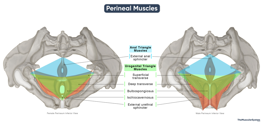
Male & Female Perineal Muscles
They are largely the same in both males and females, forming a consistent anatomical framework across the sexes. However, their size, attachment points, orientation, and specific functions may vary depending on the surrounding structures, such as the presence of the penis or vagina.
Urogenital Triangle
Seven of the eight perineal muscles are located in the urogenital triangle. It is a complex structure with several muscular and fascial layers:
- Perineal membrane: A tough fascial sheet that provides support and attachment to the perineal muscles. It divides the urogenital triangle into two compartments.
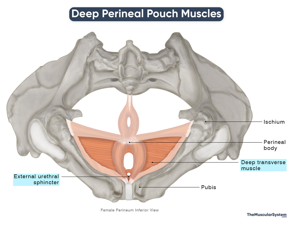
- Deep perineal pouch or space: The first of the two compartments, it lies superior to the perineal membrane. It contains two perineal muscles, along with vital structures like the urethra, vagina (in females), and bulbourethral glands (in males).
- Superficial perineal pouch or space: It lies inferior to the perineal membrane, and contains five perineal muscles, along with the erectile tissues of the penis and clitoris, and the greater vestibular glands (in females).
- Perineal fascia: It is further divided into:
- Deep perineal fascia: A fascial layer continuous with the anterior abdominal wall’s Scarpa’s fascia.
- Superficial perineal fascia: The most superficial fascial layer continuous with the anterior abdominal wall’s Camper’s fascia.
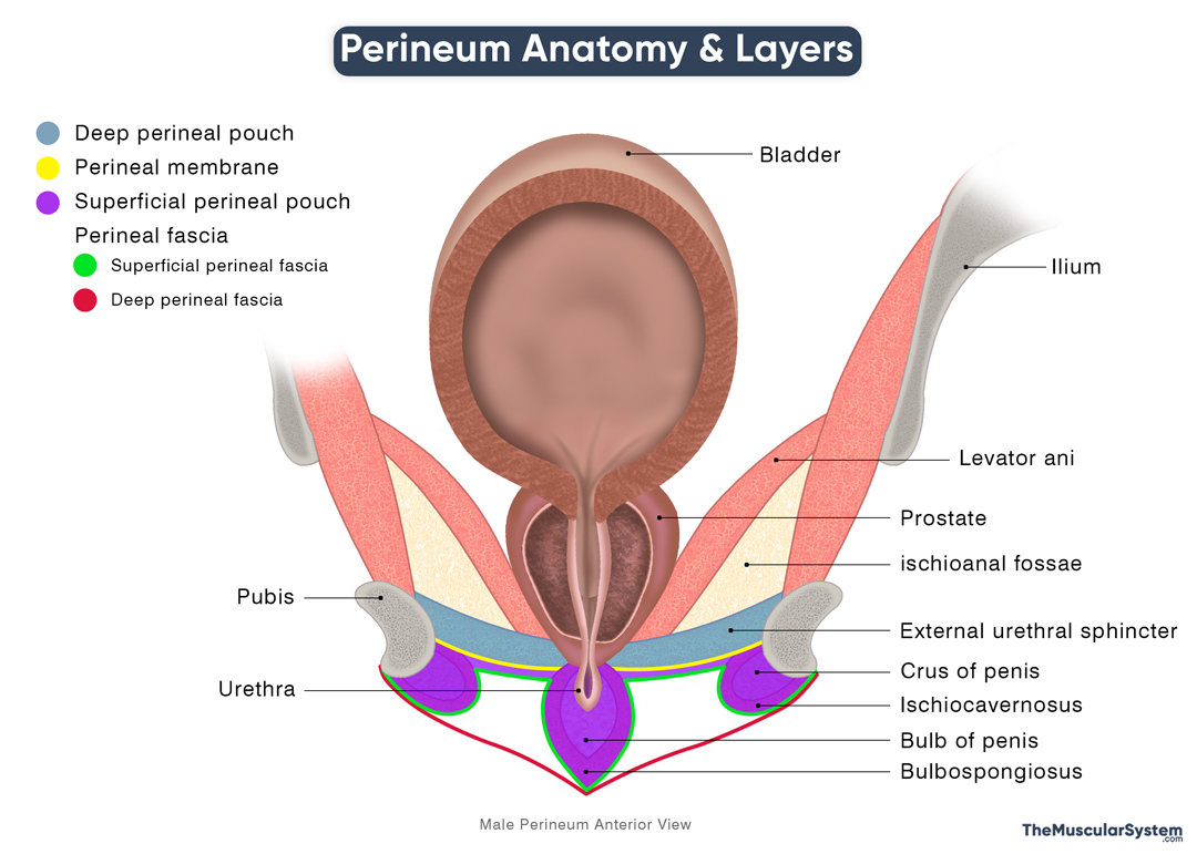
Perineum Anatomy & Layers
Names & Anatomy of Muscles in the Deep & Superficial Perineal Pouches
| Name | Origin | Insertion | Action | Innervation |
|---|---|---|---|---|
Muscles in the Deep Perineal Pouch |
||||
| Deep Transverse Perineal Muscle | Inferior ischial ramus | Contralateral DTP muscle fibers, and the perineal body | —Supporting the perineum
— Helping restore pelvic function after childbirth |
Perineal branch of the pudendal nerve (S2-S4) |
| External Urethral Sphincter | ||||
| — In males | Ischial rami, inferior pubic rami, and adjacent fascia | Contralateral EUS muscle fibers | ||
| — In females (has three distinct parts) | Same | 1) Innermost part: Forms a true muscular ring around the urethra with no distinct point of “insertion”
2) Urethral compressor: Loops around the urethra, blending with fibers from the contralateral muscle 3) Urethrovaginal sphincter: Encircles the urethra and vagina, blending with the contralateral fibers. |
Providing voluntary control over the urethral opening | Perineal branch of the pudendal nerve (S2-S4) |
Muscles in the Superficial Perineal Pouch |
||||
| Superficial Transverse Perineal | Anterior surface of the ischial tuberosity | Contralateral STP muscle fibers, and the perineal body | — Supporting the perineum
— Helping restore pelvic function following childbirth |
Perineal branch of the pudendal nerve (S2-S4) |
| Bulbospongiosus | ||||
| — In males | Penile median raphe, and perineal body | Perineal membrane, bulb of penis, penile median raphe, penile shaft | — Draining out the last of the urine from the urethra
— Assisting with penile erection |
Perineal branch of the pudendal nerve (S2-S4) |
| — In females | Perineal body | Clitoral corpora cavernosa | — Draining urine
— Assisting with clitoral erection, and vaginal tightening |
Same |
| Ischiocavernosus | ||||
| — In males | Ischial tuberosity and ischial ramus | Crus of the penis | Maintaining erection of the penis | Perineal branch of the pudendal nerve (S2-S4) |
| — In females | Same | Crus of the clitoris | Maintaining erection of the clitoris | Same |
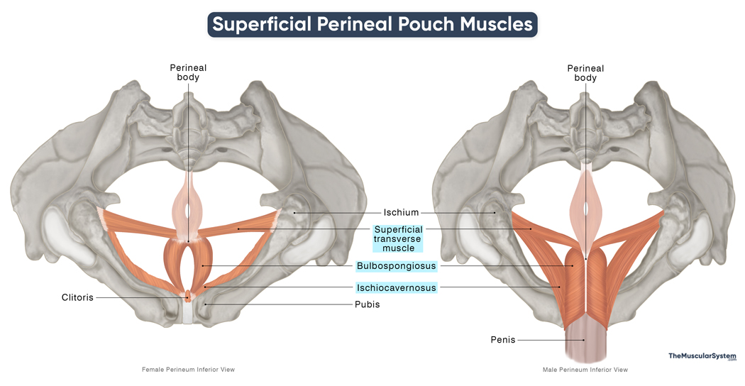
Superficial Perineal Pouch Muscles
Deep Superficial Perineal Pouch Muscles
Anal Triangle
The anal or anorectal triangle is much less complex than the urogenital triangle, with the main structures including the:
- The anus, or the opening of the anal canal.
- The ischioanal fossae, two fat-filled spaces on either side of the anal canal that help with defecation
- The anal triangle contains perineal, pelvic, and hip muscles, including the external anal sphincter, the levator ani group, the coccygeus (pelvic floor muscles), and the obturator internus (hip muscle).
Name & Anatomy of Perineal Muscles in the Anal Triangle
| Name | Attachment | Action | Innervation |
| External anal sphincter | 1) Deep part: At the back of the anococcygeal raphe
2) Superficial part: The perineal body at the front, and the coccyx at the back 3) Subcutaneous part: Forms a muscular ring around the lowest part of the anal canal, with no distinct points of attachment |
Providing voluntary control over the anal canal and opening | Perineal and inferior rectal branches of the pudendal nerve (S2-S4) |
Note: The lists above include only the skeletal muscles of the perineum, which are under somatic (voluntary) control via the pudendal nerve. It excludes the internal urethral sphincter, and internal anal sphincter, which are smooth muscles regulated by the autonomic nervous system, and therefore not considered part of the voluntary perineal musculature.
References
- Perineum: ClevelandClinic.org
- The Perineum: TeachMeAnatomy.info
- Anatomy, Abdomen and Pelvis, Perineal Body: NCBI.NLM.NIH.gov
- Perineal Region: Kenhub.com
- Muscles of the Pelvis and Perineum: https://Medicine.UAMS.edu
- Anatomy, Abdomen and Pelvis: Superficial Perineal Space: NCBI.NLM.NIH.gov
- Deep perineal pouch: Radiopaedia.org


