

Bulbospongiosus
By
Della Barnes, an MS Anatomy graduate, blends medical research with accessible writing, simplifying complex anatomy for a better understanding and appreciation of human anatomy.
Last updated:
20/06/2025Della Barnes, MS Anatomy
UX/UI Designer at - AdobeDella Barnes, an MS Anatomy graduate, blends medical research with accessible writing, simplifying complex anatomy for a better understanding and appreciation of human anatomy.
What is the Bulbospongiosus
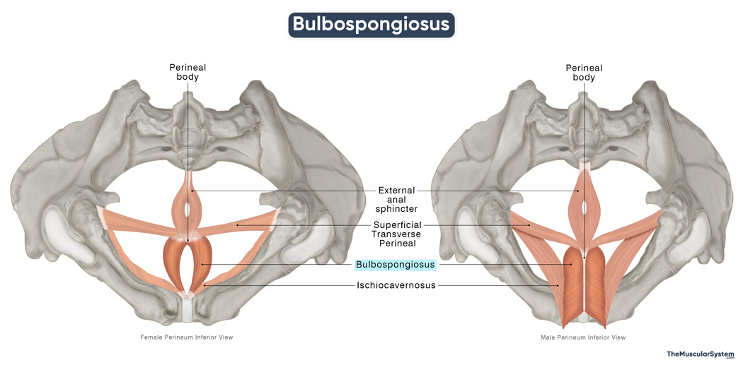
The bulbospongiosus, or bulbocavernosus (in older texts), is a paired muscle found at the base of the genitals in both males and females. It is one of the 3 muscles belonging to the superficial perineum pouch, the others being the ischiocavernosus and superficial transverse perineal.
Its attachments, functions, and appearance vary slightly in males and females. The muscle aids in urination, sexual function, and muscle control in that area.
Anatomy
Location and Attachments
| Origin | In males: The median raphe of penis and perineal body In females: The perineal body |
| Insertion | In males: The perineal membrane, bulb of the penis, median raphe of penis, and the lateral side of the penile shaft In females: The clitoral corpora cavernosa |
Origin
In both males and females, the bulbospongiosus muscle originates via tendinous fibers from the perineal body, the central fibromuscular structure in the perineum that serves as the anchoring point for several perineal muscles, including the superficial transverse perineal and external anal sphincter muscles. Some of its fibers may also blend with adjacent musculature.
In males, the muscle additionally arises from the median raphe over the bulb of the penis, sometimes referred to as the median penile raphe.
Insertion
In Males
From their central origin, the muscle fibers fan out obliquely upward, resembling the barbs of a feather. These fibers insert at different locations within the perineal region, and the muscle can be functionally divided into three parts based on their insertion points:
1. The posterior fibers form a thin muscular layer that blends seamlessly with the perineal membrane.
2. The central fibers are broader and wrap around the bulb of the penis — the bulbous end of the corpus spongiosum, which is one of the erectile tissues of the penis. Fibers from the left and right bulbospongiosus converge at the dorsal midline, contributing to the median raphe of the penis that supports and stabilizes the penial region.
3. The anterior fibers extend forward over the corpus spongiosum and insert along the lateral aspects of the penile shaft, just anterior to the ischiocavernosus muscle. These fibers commonly blend with a tendinous sheet that lies over the dorsal penile blood vessels, contributing to the structural integrity of the area.
In Females
In females, there is no median raphe of the penis, and the bulbospongiosus muscles on either side typically remain separate, without midline fusion. From their origin at the perineal body, the muscle fibers course laterally to insert into the clitoral corpora cavernosa, the sponge-like erectile tissue within the clitoris above the vaginal opening.
Relations With Surrounding Muscles and Structures
Being a muscle in the superficial perineal pouch, it is part of the muscular superficial layer of the perineum. Along with the other superficial perineal pouch muscles, the ischiocavernosus and superficial transverse perineal, the bulbospongiosus is invested within the deep perineal fascia, also called the fascia of Gallaudet.
Among the structures associated with the bulbospongiosus muscle is the tendinous perineal body, which lies anterior to it. In males, the muscle covers the bulb of the penis. In females, it lies on either side of the vaginal orifice, overlying the vestibular bulbs and partially covering the clitoral bulbs.
Function
| Action | Helping with draining out urine Aiding penile erection (in males), clitoral erection, and vaginal tightening (in females) |
General Function
- Assists in expelling the final drops of urine from the urethra after emptying the bladder.
In Males
- Aids in maintaining an erection by compressing the veins at the base of the penis, helping to trap blood within the erectile tissue.
- Contributes to ejaculation through rhythmic contractions that help propel semen through the urethra.
- Helps clear the urethra of any remaining semen.
In Females
- Tightens the vaginal opening, which can be important during activities like sex and childbirth.
- Assists in the release of lubricating fluids from the greater vestibular glands.
- Aids in clitoral erection by compressing the erectile tissue, similar to its role in males.
Innervation
| Nerve | Perineal branch of the pudendal nerve (S2-S4) |
The muscle gets innervated by the perineal nerve, a terminal branch of the pudendal nerve originating from the 2nd to 4th sacral nerve roots (S2 to S4). It is the primary nerve supply to the perineal region.
Blood Supply
| Artery | Perineal artery |
It receives blood supply from the perineal artery, which branches off from the internal pudendal artery.
References:
- Bulbospongiosus Muscle: Elsevier.com
- Bulbospongiosus Muscle: Kenhub.com
- Bulbospongiosus Muscle: Radiopaedia.org
- Bulbospongiosus Muscle: IMAIOS.com
- Bulbospongiosus Muscle: PrimaryCareNotebook.com
Della Barnes, an MS Anatomy graduate, blends medical research with accessible writing, simplifying complex anatomy for a better understanding and appreciation of human anatomy.
- Latest Posts by Della Barnes, MS Anatomy
-
Tensor Tympani
- -
Stapedius
- -
Auricularis Posterior
- All Posts


