

Lumbricals of the Foot
By
Della Barnes, an MS Anatomy graduate, blends medical research with accessible writing, simplifying complex anatomy for a better understanding and appreciation of human anatomy.
Last updated:
29/10/2025Della Barnes, MS Anatomy
UX/UI Designer at - AdobeDella Barnes, an MS Anatomy graduate, blends medical research with accessible writing, simplifying complex anatomy for a better understanding and appreciation of human anatomy.
What Are the Lumbricals
The lumbricals are a group of four small muscles located on the sole of the foot, associated with the four lateral digits. They are numbered from medial to lateral as the first to fourth lumbricals. These muscles belong to the second layer of the plantar muscles, together with the quadratus plantae.
The name “lumbricals” is derived from the Latin word “lumbricus,” meaning worm; it refers to the thin, wormlike structure of these four muscles. Still, they play an important role in the movement and coordination of the second to fifth toes.
Anatomy
Location and Attachments
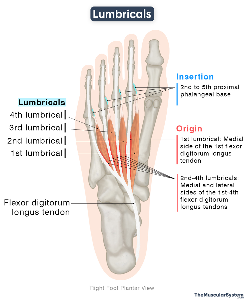
| Origin | The medial and lateral sides of the 1st to 4th flexor digitorum longus tendons |
| Insertion | The 2nd to 5th proximal phalangeal base and the attached extensor expansions |
Origin
They originate from the tendons of the flexor digitorum longus (FDL). The first lumbrical arises from the medial side of the first FDL tendon, which inserts into the second toe. The second, third, and fourth lumbricals arise from the adjacent sides of all four FDL tendons. Consequently, the first lumbrical is unipennate, whereas the remaining three are bipennate.
Because these muscles originate from the FDL tendons, they are sometimes described as accessory muscles of the FDL.
Insertion
The small muscle bellies course forward and then narrow into thin tendons as they reach the phalanges. Finally, they insert into the bases of the second to fifth proximal phalanges and their adjacent extensor expansions (or hoods).
Variations
Anatomical variations may include the absence of one or more lumbricals, the presence of additional slips, or variations in their points of insertion. Some of these variations can result in physical or functional abnormalities. For instance, the absence of one or more lumbrical muscles may lead to gait disturbances or deformities such as hammer toes and claw toes.
Relations With Surrounding Muscles and Structures
On the proximal side, the lumbrical muscles lie deep to the plantar aponeurosis and superficial to the adductor hallucis, which belongs to the third layer of the plantar muscles. All four lumbricals lie lateral to the flexor hallucis brevis. Apart from the first lumbrical, the remaining three are medial to the tendon of the flexor digitorum longus to the second toe.
On the distal side, the lumbricals pass along the plantar surface of the deep transverse metatarsal ligament, which connects the heads of the metatarsal bones and supports the metatarsophalangeal joints.
Function
| Action | Flexing the metatarsophalangeal joints, and extending the interphalangeal joints of the 2nd to 5th toes |
The lumbricals work together with the long flexors, the flexor hallucis longus, flexor digitorum longus, and extensors, the extensor hallucis longus, extensor digitorum longus of the toes, to coordinate fine movements and help maintain balance during gait. Each lumbrical acts on its corresponding toe — the first on the second toe, the second on the third, and so on. They simultaneously produce the following movements in their respective toes:
- Flexion at the metatarsophalangeal (MTP) joints to bend the toes at their bases.
- Extension at the interphalangeal (IP) joints to keep the toes straight at their tips.
When these muscles contract medially, they also assist in adducting the toes toward the midline, helping to keep them together during walking and running.
When the feet push off the ground during walking, the lumbricals balance toe movements, preventing them from bending too far back or curling under, which avoids “clawing” and ensures a smooth, stable stride.
The lumbricals of the foot are analogous to the lumbricals of the hand, as both have the same actions on the metacarpophalangeal and interphalangeal joints of the 2nd to 5th toes/fingers.
Innervation
| Nerve | 1st lumbrical: Medial plantar nerve (S2-S3)2nd-4th lumbricals: Lateral plantar nerve (S2-S3) |
The first lumbrical receives innervation from the medial plantar nerve, the larger terminal branch of the tibial nerve. The remaining three lumbricals are innervated by the lateral plantar nerve, the smaller terminal branch. Both nerves contain fibers derived from the first and second sacral nerve roots (S1–S2).
Blood Supply
| Artery | Medial and lateral plantar arteries |
Its blood supply comes from the medial plantar artery and the lateral plantar artery, both branches of the posterior tibial artery. Additional supply comes from the four plantar metatarsal arteries arising from the plantar arch.
References
- Lumbrical Muscles of the Foot: Kenhub.com
- Lumbricals (Foot): TeachMeAnatomy.info
- Lumbrical Muscles of the Foot: Radiopaedia.org
- Lumbrical Muscles of Foot (Left): Elsevier.com
- Morphological Variations of Foot Lumbricals and Their Clinical Significance: A Cadaveric Study: PMC.NCBI.NLM.NIH.gov
Della Barnes, an MS Anatomy graduate, blends medical research with accessible writing, simplifying complex anatomy for a better understanding and appreciation of human anatomy.
- Latest Posts by Della Barnes, MS Anatomy
-
Tensor Tympani
- -
Stapedius
- -
Auricularis Posterior
- All Posts


