

Flexor Hallucis Brevis
By
Della Barnes, an MS Anatomy graduate, blends medical research with accessible writing, simplifying complex anatomy for a better understanding and appreciation of human anatomy.
Last updated:
29/10/2025Della Barnes, MS Anatomy
UX/UI Designer at - AdobeDella Barnes, an MS Anatomy graduate, blends medical research with accessible writing, simplifying complex anatomy for a better understanding and appreciation of human anatomy.
What is the Flexor Hallucis Brevis
Flexor hallucis brevis, or FHB, is a two-headed intrinsic muscle on the medial side of the foot. It belongs to the third layer of plantar muscles, along with the adductor hallucis and flexor digiti minimi brevis. As evident from the name, it is the primary muscle responsible for flexing the hallux or big toe.
Anatomy
The muscle is divided into the lateral and medial heads, with only the lateral head having a bony origin.
Location and Attachments
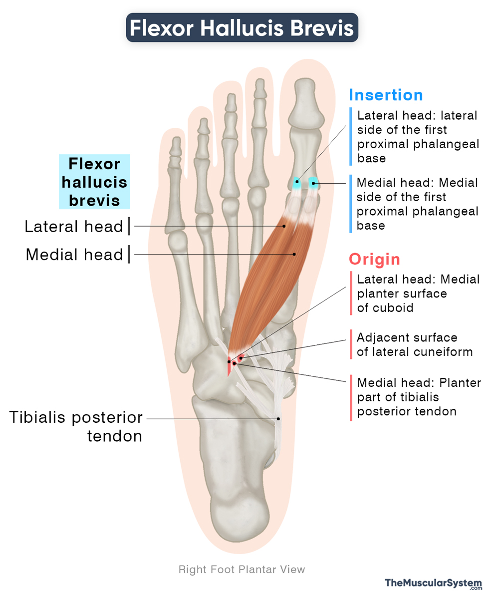
| Origin | — Lateral head: Medial plantar surface of the cuboid, and the adjacent lateral cuneiform — Medial head: Plantar part of the tibialis posterior tendon |
| Insertion | The lateral and medial sides of the proximal phalangeal base of the big toe |
Origin
The lateral head originates from the medial side of the cuboid bone’s plantar surface and the adjacent part of the lateral cuneiform. This point of origin lies just behind the groove through which the tendon of the fibularis longus muscle passes as it enters the sole of the foot.
The medial head has a tendinous origin as it rises from the plantar part of the tibialis posterior tendon, which partly inserts into the cuboid and lateral cuneiform. Some fibers of the medial head may also originate from the adjacent intermuscular septum.
Insertion
From their origin, both heads run forward to form distinct muscle bellies that narrow down to form two tendons. The lateral tendon inserts on the lateral side of the base of the big toe’s proximal phalanx, and the medial tendon inserts on the medial side of the same bone.
Flexor Hallucis Brevis Relations With Surrounding Muscles and Structures
The FHB is the most medial muscle in the third layer of the sole. It lies medial to the adductor hallucis and the first lumbrical.
As its medial and lateral tendons pass forward toward the great toe, the flexor hallucis longus tendon runs between the two. Just before their insertion into the first proximal phalanx, the medial tendon blends with the tendon of the abductor hallucis, and the lateral tendon blends with the tendon of the adductor hallucis.
Each of the two FHB tendons contains a sesamoid bone, the medial (tibial) and lateral (fibular) sesamoids, which articulate with the head of the first metatarsal. They act as small pulleys, increasing the efficiency of the two tendons during toe movement.
The medial plantar nerve lies along the medial side of the muscle.
Function
| Action | Flexing the big toe at its metatarsophalangeal joint, helping stabilize the big toe and the foot |
Flexion of the big toe
The flexor hallucis brevis primarily flexes the first metatarsophalangeal joint, bending the big toe toward the sole. This movement is essential during the pre-swing phase of walking, running, and jumping, when the toes push off the ground to help propel the body forward.
Stabilization of the big toe
Working together with the adductor hallucis and abductor hallucis, the muscle’s two tendons help stabilize the big toe. It helps the toe to maintain firm contact with the ground and stay properly aligned during movement, enhancing balance and efficiency.
Support of the medial longitudinal arch
By connecting the tarsal bones of the midfoot to the proximal phalanx of the big toe, the flexor hallucis brevis helps maintain the medial longitudinal arch. Its contraction draws these bones closer, elevating and supporting the arch during weight-bearing activities.
Antagonists
The extensor hallucis longus is the primary antagonist of the FHB, as it extends the great toe, which is the opposite action of toe flexion.
Innervation
| Nerve | Medial plantar nerve (S1-S2) |
Both heads of the muscle receive innervation from the medial plantar nerve, the larger of the two terminal branches of the tibial nerve, carrying fibers from the first and second sacral nerve roots (S1–S2).
Blood Supply
| Artery | Medial plantar artery, and first metatarsal artery |
The medial plantar artery, a branch of the posterior tibial artery, provides blood supply to the muscle. Additional blood supply comes from the first metatarsal artery, which arises from the plantar arch.
References
- Flexor Hallucis Brevis: Elsevier.com
- Flexor Hallucis Brevis Muscle: Kenhub.com
- Flexor Hallucis Brevis: TeachMeAnatomy.info
- Flexor Hallucis Brevis: IMAIOS.com
Della Barnes, an MS Anatomy graduate, blends medical research with accessible writing, simplifying complex anatomy for a better understanding and appreciation of human anatomy.
- Latest Posts by Della Barnes, MS Anatomy
-
Tensor Tympani
- -
Stapedius
- -
Auricularis Posterior
- All Posts


