

Abductor Hallucis
By
Della Barnes, an MS Anatomy graduate, blends medical research with accessible writing, simplifying complex anatomy for a better understanding and appreciation of human anatomy.
Last updated:
17/10/2025Della Barnes, MS Anatomy
UX/UI Designer at - AdobeDella Barnes, an MS Anatomy graduate, blends medical research with accessible writing, simplifying complex anatomy for a better understanding and appreciation of human anatomy.
What is the Abductor Hallucis
The abductor hallucis is a small fusiform muscle on the plantar side of the foot. It lies on the medial side of the foot, forming the fleshy lump just in front of the heel, under the foot.
The muscle forms the superficial or first layer of the plantar muscles along with the flexor digitorum brevis and abductor digiti minimi. As its name suggests, it contributes to abduction and flexion of the big toe.
Anatomy
Location and Attachments
| Origin | Medial process of calcaneal tuberosity, the flexor retinaculum, and the plantar aponeurosis |
| Insertion | Medial side of the base of the first proximal phalanx (of the big toe) |
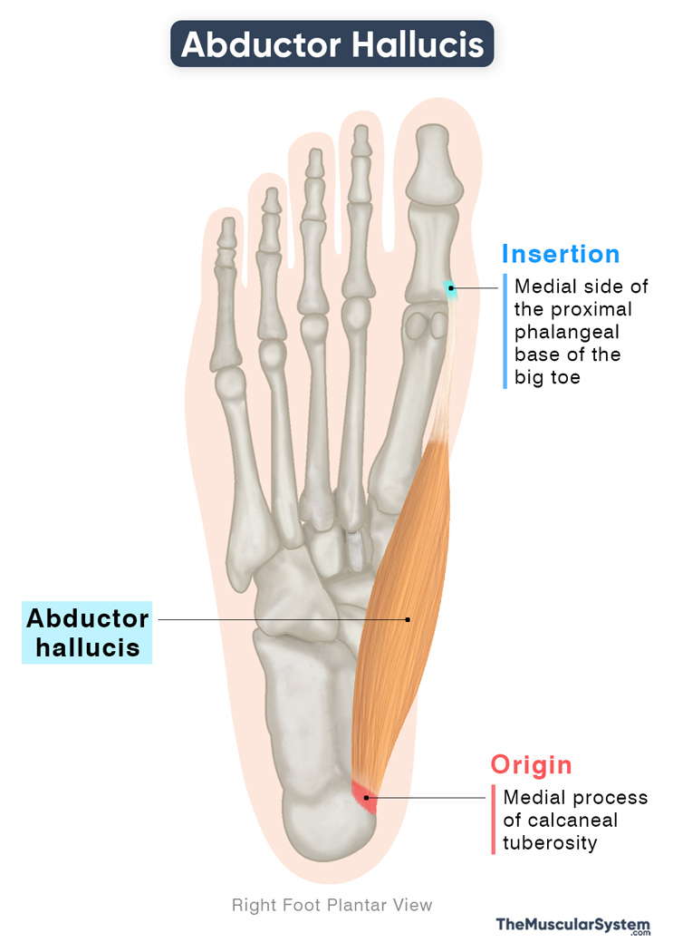
Origin
The slender intrinsic muscle originates from the medial process/tubercle of the calcaneal tuberosity on the calcaneus or heel bone. It also takes origin from the flexor retinaculum of the foot, and the plantar aponeurosis, the layer of fibrous tissue that forms the sole of the foot.
Insertion
After originating, the muscle belly courses toward the front along the medial border of the foot. As it approaches the body of the first metatarsal, it tapers into a tendon that joins the medial head of the flexor hallucis brevis. Together, they insert into the medial side of the base of the proximal phalanx of the great toe and the medial sesamoid bone associated with the first metatarsophalangeal joint.
Relations With Surrounding Muscles and Structures
The abductor hallucis is the most medial muscle of the foot, positioned along its inner border and contributing to the contour of the medial margin. It lies directly beneath the plantar aponeurosis. Deep to the muscle are the flexor digitorum longus tendon, the medial plantar artery, and the medial plantar nerve. Laterally, the abductor hallucis is related to the flexor digitorum brevis.
At its proximal attachment, the calcaneus bone and the fibers of the abductor hallucis together form an opening known as the porta pedis, a passageway through which the medial and lateral plantar nerves and vessels pass into the sole.
Function
| Action | Abduction and flexion of the big toe |
Despite its relatively small size, the abductor hallucis is a strong muscle, playing an important role in walking.
- Its primary action is to abduct the big toe at its metatarsophalangeal joint.
- Due to its attachment to the medial side of the great toe, the muscle helps keep the big toe in a central position during walking.
- It also assists the flexor hallucis longus and flexor hallucis brevis in flexing the big toe.
- Due to its attachment from the heel bone to the first proximal phalanx, it also supports the medial longitudinal arch of the foot, helping maintain balance and stability while walking.
Injury or weakness of this muscle may lead to hallux valgus, a deformity where the big toe deviates toward the other toes.
The abductor hallucis in the foot is functionally similar to the opponens pollicis in the hand. Both are intrinsic muscles on the medial side of the foot or hand, and act on the first digit, the big toe or thumb. While the big toe’s muscle provides stability during standing and walking, the thumb’s muscle enhances precision and fine movement.
Antagonists
The adductor hallucis acts as the primary antagonist of this muscle, since it adducts the big toe, performing the opposite action of the abductor hallucis.
Innervation
| Nerve | Medial plantar nerve (S1-S3) |
The muscle is supplied by the medial plantar nerve, which enters through its upper border. The medial plantar nerve is the larger terminal branch of the tibial nerve, and it originates from the first to third sacral nerve roots (S1-S3).
Blood Supply
| Artery | Medial plantar artery and first plantar metatarsal artery |
The muscle receives its blood supply primarily from the medial plantar artery, a branch of the posterior tibial artery. The first plantar metatarsal artery, a branch of the lateral plantar artery, also contributes to its blood supply.
References
- Abductor Hallucis: TeachMeAnatomy.info
- Abductor Hallucis Muscle: Kenhub.com
- Abductor Hallucis Muscle: Radiopaedia.org
- Abductor Hallucis: Elsevier.com
Della Barnes, an MS Anatomy graduate, blends medical research with accessible writing, simplifying complex anatomy for a better understanding and appreciation of human anatomy.
- Latest Posts by Della Barnes, MS Anatomy
-
Tensor Tympani
- -
Stapedius
- -
Auricularis Posterior
- All Posts


