

Posterior Compartment of the Leg
By
Della Barnes, an MS Anatomy graduate, blends medical research with accessible writing, simplifying complex anatomy for a better understanding and appreciation of human anatomy.
Last updated:
27/09/2025Della Barnes, MS Anatomy
UX/UI Designer at - AdobeDella Barnes, an MS Anatomy graduate, blends medical research with accessible writing, simplifying complex anatomy for a better understanding and appreciation of human anatomy.
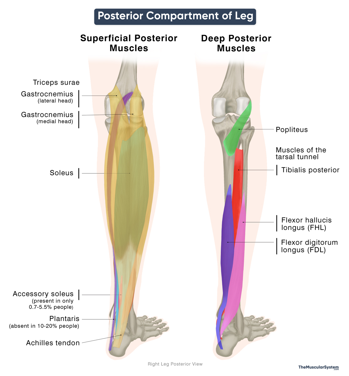
The posterior compartment of the leg is a fascial compartment that contains the muscles at the back of the lower leg. It is divided into superficial and deep posterior compartments, separated by the transverse intermuscular septum. The tibia, fibula, and interosseous membrane separate it from the anterior compartment, while the posterior intermuscular septum separates it from the lateral compartment.
There are seven muscles in the posterior lower leg, divided between the superficial and deep compartments. These muscles are responsible for plantarflexion, toe flexion, and inversion, functions essential for walking and maintaining foot stability.
Muscles in the Posterior Compartment and Their Anatomy
The superficial group consists of three muscles that act together to produce plantarflexion at the ankle. The deep group has four muscles, three of which pass through the tarsal tunnel, contributing to plantarflexion and toe flexion.
The following table summarizes the muscles of this compartment, along with their attachments and functions:
| Name | Origin | Insertion | Function |
|---|---|---|---|
| Superficial Posterior Leg Muscles | |||
| Triceps surae* | |||
| — Soleus | — Posterior surface of the tibia (soleal line and middle third of the medial border) — Posterior surface of the fibula (head and upper one-fourth of the shaft) | Posterior surface of the calcaneus, via the Achilles tendon | Plantar flexion at the ankle joint |
| — Gastrocnemius | — Lateral head: Posterior surface of the lateral condyle of the femur — Medial head: Posterior surface of the medial condyle and the popliteal surface of the femur | Posterior surface of the calcaneus, via the Achilles tendon | Plantar flexion and knee flexion |
| Plantaris (may be absent in 10–20% of the population) | Lower part of the lateral supracondylar ridge of the femur | Posterior surface of the calcaneus, usually via the Achilles tendon | — Plantar and knee flexion (weak) — May also play a role in proprioception due to the high density of muscle spindles |
| Deep Posterior Leg Muscles | |||
| Popliteus | Lateral condyle of the femur | Posterior surface of the proximal tibia | Unlocks the knee by rotating the tibia/femur to assist flexion of the fully extended knee joint. |
| Flexor hallucis longus (FHL)# | Posterior surface of the distal two-thirds of the fibula | Distal phalangeal base of the big toe (plantar surface) | Flexing the big toe at all its joints, and helping with plantar flexion |
| Flexor digitorum longus (FDL)# | Posterior surface of the tibial body | Distal phalangeal bases of the 2nd to 5th toes (plantar surface) | Flexing the 2nd to 4th toes at all their joints, and helping with plantar flexion |
| Tibialis posterior# | — Posterior surface of the upper half of the tibia — Upper two-thirds (2/3) of the medial-posterior fibula — Posterior surface of the interosseous membrane | — Tuberosity of the navicular bone — Plantar surfaces of all three cuneiforms — Plantar surface of the cuboid bone — Bases of the second to fourth metatarsals — Sustentaculum tali of the calcaneus bone | Foot inversion, plantarflexion (ankle), stabilization of the medial longitudinal arch |
*The triceps surae is a three-headed muscle group that forms the calf, composed of the two-headed gastrocnemius and the soleus. Their common tendon, the Achilles tendon, is the strongest and thickest tendon in the human body. In some individuals (0.7% to 5.5%), an additional muscle, the accessory soleus, also inserts into the Achilles tendon.
#The distal tendons of these three muscles pass through the tarsal tunnel. From anterior to posterior, the order is tibialis posterior, FDL, posterior tibial artery and vein, tibial nerve, and FHL. A common mnemonic for recalling this sequence is “Tom, Dick And Very Nervous Harry.”
Nerve and Blood Supply
Being in the same compartment, all posterior leg muscles share the nerve supply. However, the superficial and deep groups receive blood from different sources.
Innervation: Tibial nerve (L4-S3)
Blood supply:
- Superficial group: Mainly the sural artery, a branch of the popliteal artery, with additional supply from the femoral and posterior tibial arterial branches.
- Deep group: Branches of the popliteal, fibular, and posterior tibial arteries.
References
- Muscles in the Posterior Compartment of the Leg: TeachMeAnatomy.info
- Anatomy, Bony Pelvis and Lower Limb: Leg Posterior Compartment: NCBI.NLM.NIH.gov
- Deep posterior muscles of the leg: Kenhub.com
- Posterior compartment of leg: IMAIOS.com
Della Barnes, an MS Anatomy graduate, blends medical research with accessible writing, simplifying complex anatomy for a better understanding and appreciation of human anatomy.
- Latest Posts by Della Barnes, MS Anatomy
-
Tensor Tympani
- -
Stapedius
- -
Auricularis Posterior
- All Posts


