Plantaris
Last updated:
04/09/2025Della Barnes, an MS Anatomy graduate, blends medical research with accessible writing, simplifying complex anatomy for a better understanding and appreciation of human anatomy.
What is the Plantaris
The plantaris is a long, narrow muscle located behind the knee, extending down the calf to the heel. It forms part of the superficial posterior compartment of the leg along with the gastrocnemius and soleus, assisting them in actions such as plantar flexion and knee flexion. However, the muscle is sometimes considered vestigial, as it may be absent in certain individuals.
Plantaris gets its name from its insertion into the plantar aponeurosis in many mammals, though it does not reach it in humans.
Anatomy
Location and Attachments
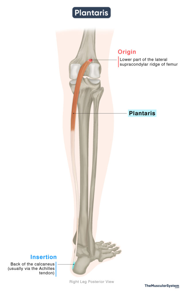
| Origin | Lower part of the lateral supracondylar ridge of the femur |
| Insertion | Back of the calcaneus (usually via the Achilles tendon) |
Origin
The muscle originates from the lower end of the lateral supracondylar ridge on the femur, just superior to the origin of the lateral head of the gastrocnemius. Sometimes, the attachment may extend to the oblique popliteal ligament. The fusiform belly of the muscle is quite short by itself, measuring about 5–10 cm in length. It quickly tapers into a long, thin tendon.
Insertion
The tendon then descends medially to pass to the back of the knee joint and then travels down the calf region towards the heel. It typically blends with the Achilles tendon, which is the common tendon for all the superficial muscles in the posterior compartment. The Achilles tendon then inserts into the back of the calcaneus, or heel bone. Sometimes, the tendon may travel medial to the Achilles tendon instead of blending with it, inserting into the medial side of the calcaneus.
Because of its delicate, cord-like form, the plantaris tendon may often be mistaken for a nerve during dissections, especially by new students, which is why it’s sometimes known as the “freshman’s nerve.”
Relations With Surrounding Muscles and Structures
Within the superficial posterior compartment of the leg, the plantaris muscle lies deep to the medial head of the gastrocnemius and is also related laterally to its lateral head. As the muscle tapers into its slender tendon, this relationship is maintained, with the tendon descending along the medial border of the gastrocnemius down the posterior medial side of the lower leg.
It may help form the lower boundary of the popliteal fossa, a diamond-shaped hollow at the back of the knee, together with the gastrocnemius.
The soleus, the deeper muscle of the superficial posterior compartment, and the popliteus from the deep posterior compartment are located beneath the plantaris.
Variation
Studies show that the plantaris muscle is highly variable and may even be absent in 10–20% of people. When present, its size and shape can differ significantly. For example, the tendon may divide the muscle belly into two parts, or there may be two thin tendons instead of one.
Function
| Action | Plantar and knee flexion (weak) |
Role in ankle and knee movement
Because the muscle crosses both the knee and ankle joints, it can contribute to movements at each of these joints. However, its tendon is very small and slender, which limits its ability to generate force. For this reason, the plantaris is not considered a prime mover. Instead, it assists the gastrocnemius, soleus, and other muscles in plantar flexion and in flexing the knee.
Because the muscle contributes so little to movement, its long tendon can be used for muscle grafting with no loss of function.
Possible Role in Proprioception
Interestingly, the muscle contains a high density of muscle spindles, specialized sensory receptors that detect stretch and provide information about limb position. This suggests it may play an important role in proprioception, helping nearby muscles coordinate movement and maintain posture. However, this role has not been fully proven, since individuals who lack the muscle do not appear to lose proprioceptive ability.
Antagonists
The plantaris is too weak to have a true antagonist of its own. However, its actions, plantar flexion and knee flexion, are opposed by the ankle dorsiflexors, mainly tibialis anterior, and by the knee extensors, the quadriceps femoris.
Innervation
| Nerve | Tibial nerve (S1, S2) |
Innervation to the muscle comes from the tibial nerve, which rises from the first and second sacral nerve roots. It is a direct branch of the sciatic nerve.
Blood Supply
| Artery | Sural artery |
The muscle is primarily supplied by the sural artery, with additional supply from the superior lateral genicular artery, both arising from the popliteal artery.
References
- Plantaris Muscle: Radiopaedia.org
- Plantaris Muscle: Elsevier.com
- Plantaris Muscle: Kenhub.com
- Plantaris Muscle: IMAIOS.com
- Plantaris: TeachMeAnatomy.info
Della Barnes, an MS Anatomy graduate, blends medical research with accessible writing, simplifying complex anatomy for a better understanding and appreciation of human anatomy.
- Latest Posts by Della Barnes, MS Anatomy
-
Tensor Tympani
- -
Stapedius
- -
Auricularis Posterior
- All Posts


