

Gastrocnemius
By
Della Barnes, an MS Anatomy graduate, blends medical research with accessible writing, simplifying complex anatomy for a better understanding and appreciation of human anatomy.
Last updated:
10/10/2025Della Barnes, MS Anatomy
UX/UI Designer at - AdobeDella Barnes, an MS Anatomy graduate, blends medical research with accessible writing, simplifying complex anatomy for a better understanding and appreciation of human anatomy.
What is the Gastrocnemius
The gastrocnemius (pronunciation: GAS-trok-NEE-mee-us) is a large, two-headed, fusiform muscle that makes up the bulk of the calf. It extends from the knee to the back of the heel and forms part of the superficial posterior compartment of the lower leg, together with the soleus.
The name comes from the Greek words gaster (belly) and kneme (leg), which together mean “the belly of the leg.”
Functionally, the gastrocnemius is a primary muscle responsible for plantar flexion of the foot and also contributes to knee movement. As a result, it plays a key role in essential activities such as walking, running, and jumping.
Anatomy
The two heads of the gastrocnemius are named for the side of the leg they arise from, but they share the same point of insertion.
Location and Attachments
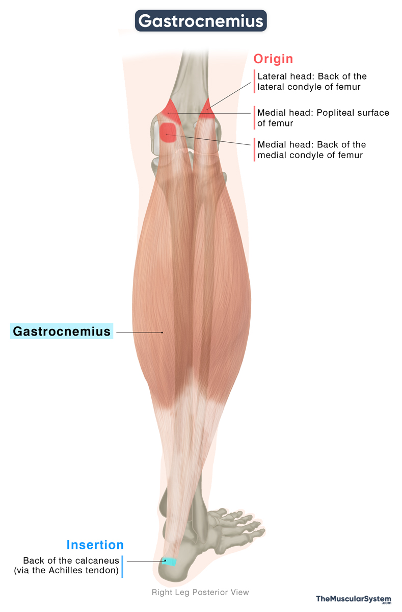
| Origin | Lateral head: The posterior surface of the lateral condyle of the femur Medial head: The posterior surface of the medial condyle and the popliteal surface of the femur |
| Insertion | Posterior surface of the calcaneus, via the Achilles tendon |
Origin
The lateral head of the gastrocnemius originates from the posterior aspect of the lateral condyle of the femur. The medial head arises from the posterior aspect of the medial condyle of the femur and extends onto the adjacent region of the popliteal surface on the distal femoral shaft.
Additionally, some fibers of both heads originate from the aponeurosis of the knee joint capsule.
Insertion
The two heads unite to form a single fleshy belly at the lower border of the popliteal fossa. This belly makes up most of the calf on the back of the leg, and its shape can be clearly seen through the skin. The muscle fibers extend roughly to the middle of the calf, where they gradually taper into a broad aponeurosis.
As it descends, this aponeurosis narrows and merges with the aponeurotic fibers of the soleus and plantaris (when present), both located in the superficial posterior compartment. Together, they give rise to the Achilles tendon, which inserts on the posterior surface of the calcaneus or heel bone in the foot.
Relations With Surrounding Muscles and Structures
Muscular relations
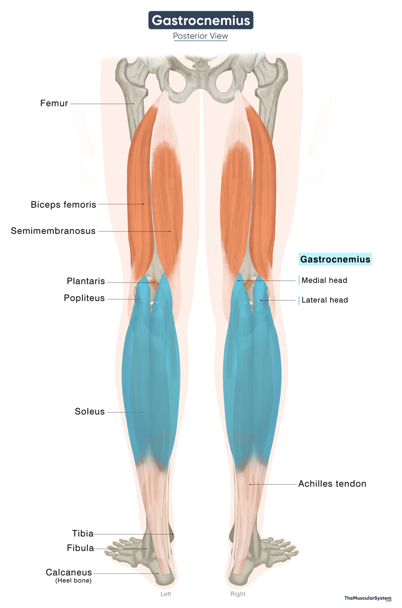
The gastrocnemius is the most superficial muscle in the posterior compartment of the leg. Deep to it lies the soleus, and together these two muscles form the three-headed triceps surae. The slender plantaris muscle runs between the bellies of the gastrocnemius and soleus.
From the deep posterior compartment, the popliteus lies deep to the proximal part of the gastrocnemius. Proximally, the muscle is also related to the hamstrings, as the tendon of the biceps femoris partly overlaps the lateral head, while the semimembranosus partly covers the medial head. Where the two heads join, they form the lower boundary of the popliteal fossa.
Nerves, blood vessels, and other structures
On the surface, the small saphenous vein and sural nerve descend along the muscle, separated from it only by the deep fascia of the leg. The common fibular nerve crosses over the lateral head as it passes between the gastrocnemius and biceps femoris. Deep to the muscle lie the popliteal artery, vein, and tibial nerve.
There is a small bursa at the origin of the medial head, near the medial femoral condyle. It reduces friction and may communicate with the semimembranosus bursa. Clinically, it is significant because enlargement of this bursa can give rise to a Baker’s cyst.
Variation
Studies show that in about 10% individuals, there is a third head of the muscle, often called the gastrocnemius tertius. It typically arises from the back of the femur and blends with either the lateral or the medial head.
Another variation is the fabella, a small sesamoid bone that sometimes develops in the tendon of the lateral head. This little bone is present in roughly 10–30% of people and may help provide extra stability to the knee joint.
Function
| Action | Plantar flexion and knee flexion |
It is a powerful muscle that produces plantar flexion of the foot at the ankle (talocrural) joint and also contributes to flexion of the leg at the knee joint. As part of the triceps surae, it works with the soleus to provide the main force for plantar flexion and is essential for propulsion in walking, running, and jumping.
During gait, this muscle is particularly active in the late stance phase (push-off), where it generates forward propulsion of the body. Unlike the soleus, which plays a greater role in maintaining upright posture, the gastrocnemius is especially engaged during fast and forceful movements of the lower limb, such as sprinting or sudden directional changes. This functional distinction reflects its predominance of type II (fast-twitch) muscle fibers, which are adapted for rapid, powerful contractions rather than sustained activity.
Antagonists
The tibialis anterior serves as the main antagonist of the gastrocnemius, countering its plantar flexion with dorsiflexion at the ankle joint.
Innervation
| Nerve | Tibial nerve (S1, S2) |
Innervation to the muscle comes from the tibial nerve, which originates from the anterior rami of the first and second sacral spinal nerves. The tibial nerve is one of the primary branches of the sciatic nerve.
Blood Supply
| Artery | Sural artery |
Both heads of the muscle recieve blood supply from the lateral and medial branches of the sural artery, which itself is a branch of the popliteal artery.
References
- Anatomy, Bony Pelvis and Lower Limb, Gastrocnemius Muscle: NCBI.NLM.NIH.gov
- Gastrocnemius: HealthLine.com
- Gastrocnemius Muscle: Kenhub.com
- Gastrocnemius Muscle: Radiopaedia.org
- Gastrocnemius (Left): Osmosis.org
- Gastrocnemius Muscle: Elsevier.com
- Gastrocnemius: TeachMeAnatomy.info
Della Barnes, an MS Anatomy graduate, blends medical research with accessible writing, simplifying complex anatomy for a better understanding and appreciation of human anatomy.
- Latest Posts by Della Barnes, MS Anatomy
-
Tensor Tympani
- -
Stapedius
- -
Auricularis Posterior
- All Posts


