Semimembranosus
Last updated:
09/08/2025Della Barnes, an MS Anatomy graduate, blends medical research with accessible writing, simplifying complex anatomy for a better understanding and appreciation of human anatomy.
What is the Semimembranosus
The semimembranosus is a large muscle located on the medial posterior side of the thigh, running from the hip joint to the knee joint. It is part of the posterior compartment of the thigh and forms the hamstring group along with the semitendinosus and biceps femoris muscles. As a hamstring muscle, it plays a key role in flexing the lower leg at the knee.
The muscle is named for its broad, membranous tendon of origin, which resembles an aponeurosis.
Anatomy
Location and Attachments
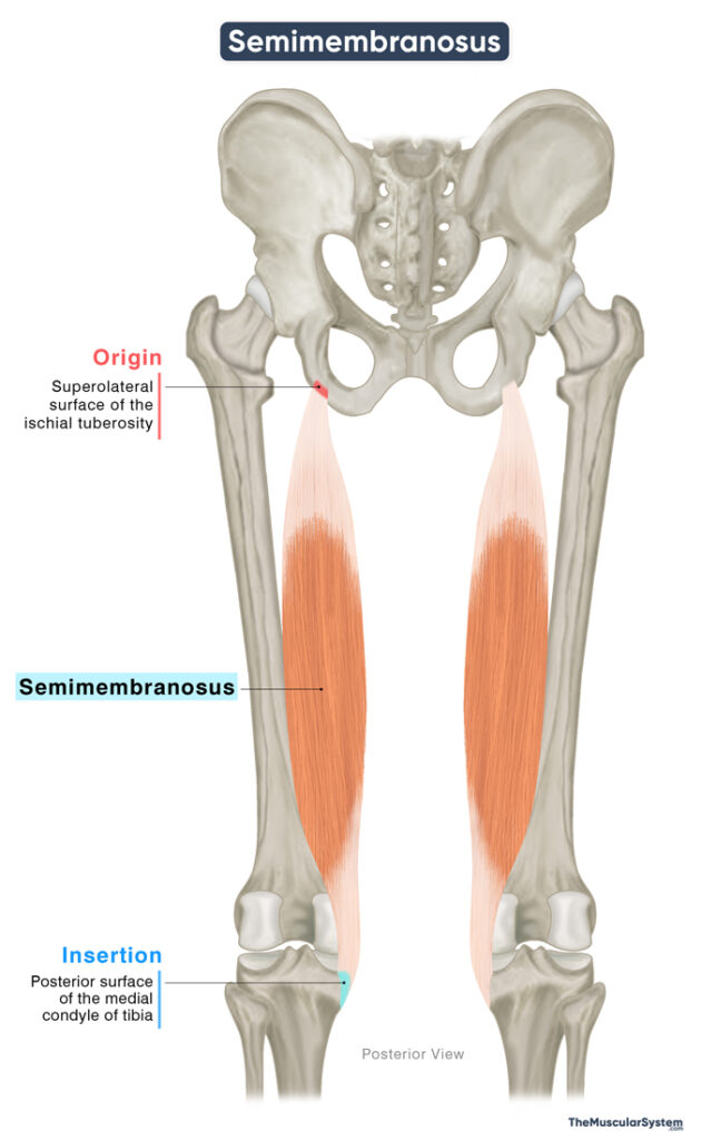
| Origin | Upper lateral surface of the ischial tuberosity |
| Insertion | Posterior surface of the medial condyle of tibia |
Origin
The muscle arises as a thick tendon from the upper and outer surface of the ischial tuberosity. This tendon expands into a membranous sheet forming the superior part of the muscle, which then gives rise to the fleshy muscle belly. The muscle belly descends toward the upper-mid thigh, converging to form the long inserting tendon.
Insertion
The tendon extends downward toward the knee joint, branching into smaller tendons that insert into different points.
- The largest part of the muscle inserts into the medial condyle of the tibia, making this the primary point of insertion.
- A second, rather prominent part of the tendon ascends laterally, contributing to the oblique popliteal ligament, also known as the posterior ligament, which then inserts into the back of the lateral condyle of the femur.
- A third portion contributes to the popliteal fascia, the deep fascia forming the roof of the popliteal fossa.
- A few fibers also insert into the medial collateral ligament, one of the largest ligaments in the knee joint.
Relations With Surrounding Muscles and Structures
Proximal Side
The point of origin of the semimembranosus muscle lies just slightly superior and lateral to the origin of the semitendinosus and the long head of the biceps femoris. Among the three hamstring muscles, the semimembranosus lies the deepest and most medial as it descends the thigh.
In the upper thigh, the gluteus maximus muscle overlies the posterior surface of the semimembranosus. As it travels downward, it lies deep to the adductor magnus and medial to the biceps femoris.
The sciatic nerve, the largest nerve in the body, courses lateral to the semimembranosus as it descends toward the knee and enters the popliteal fossa. At the level of the knee, the semimembranosus helps form the superomedial boundary of the popliteal fossa, along with the semitendinosus.
Distal Side
As the semimembranosus crosses the posteromedial aspect of the knee joint, it lies medial to the medial head of the gastrocnemius. The gracilis muscle runs more superficially and medially in the distal part of the thigh.
The U-shaped semimembranosus bursa surrounds the semimembranosus tendon at the knee joint, preventing friction between the muscle and the medial tibial plateau, the top surface of the tibia. The bursa also separates the muscle from the semitendinosus, the medial head of the gastrocnemius, and the posterior cruciate ligament.
Function
| Action | Flexing the leg at the knee joint and extending the thigh at the hip joint |
As part of the hamstring group, the semimembranosus spans the hip and knee joints, playing a key role in moving both. It works with the other two hamstring muscles to perform the following actions.
Knee flexion: The muscle contributes to flexing the knee, which means bending the knee joint that allows movements like walking and running.
Medial rotation of the tibia: When the knee is flexed, the semimembranosus aids in the medial (inward) rotation of the tibia at the knee joint. This action allows the toes to point toward the other foot.
Hip extension: It helps extend the thigh at the hip joint, working with the other hamstring muscles to pull the torso into an upright position, like when standing up.
Antagonists
The quadriceps muscles, rectus femoris, vastus lateralis, vastus intermedius, and vastus medialis, are antagonistic to the semimembranosus. While the semimembranosus flexes the knee and extends the hip, the quadriceps extend the knee and flex the hip.
Innervation
| Nerve | Tibial nerve (L5-S2) |
Since it is a hamstring muscle, its innervation comes from the tibial nerve, arising from the fifth lumbar, and the first and second sacral nerve roots (L5, S1, S2). It is a branch of the sciatic nerve.
Blood Supply
| Artery | Perforating arteries |
The primary blood supply to the semimembranosus comes from the perforating arteries, which originate from the profunda femoris artery.
Additionally, the inferior gluteal artery, a branch of the internal iliac artery, supplies the proximal part of the muscle. The muscular branches of the popliteal artery also contribute to the muscle’s vascular supply.
References
- Semimembranosus Muscle: Elsevier.com
- Semimembranosus Muscle: IMAIOS.com
- Semimembranosus Muscle: Kenhub.com
- Anatomy, Bony Pelvis and Lower Limb, Hamstring Muscle: NCBI.NLM.NIH.gov
- Semimembranosus: Rad.UW.edu
- Semimembranosus Muscle: Radiopaedia.org
Della Barnes, an MS Anatomy graduate, blends medical research with accessible writing, simplifying complex anatomy for a better understanding and appreciation of human anatomy.
- Latest Posts by Della Barnes, MS Anatomy
-
Infrahyoid Muscles
- -
Omohyoid
- -
Sternohyoid
- All Posts


