

Rectus Femoris
By
Della Barnes, an MS Anatomy graduate, blends medical research with accessible writing, simplifying complex anatomy for a better understanding and appreciation of human anatomy.
Last updated:
14/08/2025Della Barnes, MS Anatomy
UX/UI Designer at - AdobeDella Barnes, an MS Anatomy graduate, blends medical research with accessible writing, simplifying complex anatomy for a better understanding and appreciation of human anatomy.
What is the Rectus Femoris
The rectus femoris is a relatively large fusiform muscle located at the front of the thigh. It belongs to the quadriceps femoris group of muscles along with vastus medialis, vastus intermedius, and vastus lateralis.
One unique feature of the muscle is its bipennate arrangement, in which the muscle fibers are oriented in two directions, resembling the structure of a feather’s vane. It spans the distance between the hip and knee joints, and plays an important role in the movement of both, especially in forceful extension of the knee, earning it the name “kicking muscle.”
Anatomy
Location and Attachments
| Origin | Direct head: Anterior inferior iliac spine (AIIS) Indirect head: Supraacetabular groove |
| Insertion | — The base of the patella (kneecap) via the quadriceps femoris tendon — The tibial tuberosity via the patellar ligament |
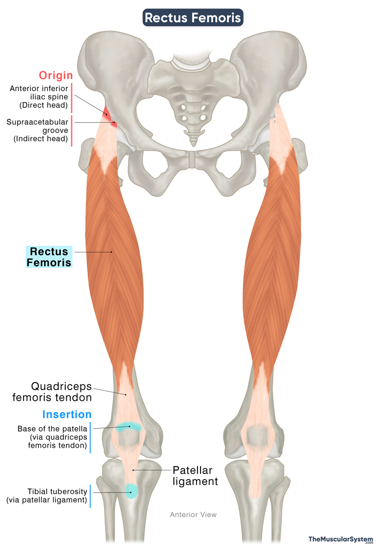
Origin
The Rectus Femoris muscle originates from two points, via two heads. The direct or straight head rises from the anterior inferior iliac spine (AIIS). The indirect or reflected head originates from the supraacetabular groove. It is the bony ridge or groove just above the acetabulum, the cup-shaped articular surface of the hip bone where the femoral head attaches to form the hip joint.
The tendons from both origins course downward, converging at an acute angle into a single tendon from where they thicken into the muscle belly with the bipennate orientation of the fibers.
Insertion
The muscle belly courses almost vertically downward toward the knee joint as the muscle fibers narrow and become tendinous. Near the knee joint, it merges with the distal tendons of the other three quadriceps muscles to form a flattened common tendon known as the quadriceps tendon. It then inserts into the base of the patella bone or kneecap via the patellar tendon.
From the apex of the patella, some part of the tendon continues downward as the patellar ligament, and attaches to the tibial tuberosity, establishing a connection between the quadriceps muscles and the tibia through the patella.
Relations With Surrounding Muscles and Structures
It is the most superficial muscle in the anterior compartment of the thigh. So, all other muscles and structures in this compartment lie deep to it, including the three vastus muscles of the quadriceps group, the lateral circumflex femoral artery, and some femoral nerve branches. Among these, the rectus femoris directly overlies the vastus intermedius, while the vastus lateralis and vastus medialis lie lateral and medial to it, respectively.

At its proximal end near the origin, the sartorius muscle, originating from the anterior superior iliac spine, crosses obliquely and lies superficially over the rectus femoris. The tensor fasciae latae lies lateral and superficial to it. The rectus femoris itself lies superficial to the iliofemoral ligament, a strong band reinforcing the anterior aspect of the hip joint capsule.
Function
| Action | Extending the knee joint and flexing the hip joint |
The muscle has two primary functions. As part of the quadriceps, the rectus femoris is one of the primary muscles that extend the leg at the knee joint. Additionally, it works alongside the ilipsoas and sartorius muscles to flex the thigh at the hip joint.
Active and Passive Insufficiencies of the muscle
Active insufficiency is where a muscle that spans multiple joints can’t produce full force because it’s already shortened or contracted across those joints.
For example, the rectus femoris, which crosses both the hip and knee joints, can experience active insufficiency when trying to extend the knee while the hip is already flexed (and vice versa). If you’re sitting in a chair, the hip is flexed and the rectus femoris is already shortened at the hip. In this position, it has limited ability to generate force to straighten the knee because it’s already contracted.
Passive insufficiency occurs when a multi-joint muscle, like the rectus femoris, is stretched over all joints it crosses, limiting range of motion. For the rectus femoris, this happens when the hip is extended and the knee is fully flexed, causing the muscle to reach its maximal length and restricting further movement.
Antagonists
The three hamstring muscles, semimembranosus, semitendinosus, and biceps femoris, act as antagonists to the rectus femoris, as their primary actions are knee flexion and hip extension, which oppose the actions of the rectus femoris.
Innervation
| Nerve | Femoral nerve (L2-L4) |
The rectus femoris muscle is innervated by the femoral nerve, which originates from the 2nd to 4th lumbar spinal nerve roots (L2 to L4). It is the largest branch rising from the lumbar plexus.
Blood Supply
| Artery | Descending branch of the lateral circumflex femoral artery |
The rectus femoris muscle is primarily supplied by the descending branch of the lateral circumflex femoral artery, a branch of the deep femoral artery.
References
- Anatomy, Abdomen and Pelvis, Rectus Femoris Muscle: NCBI.NLM.NIH.gov
- Rectus Femoris Muscle: Radiopaedia.org
- Rectus Femoris: TeachMeAnatomy.info
- Rectus Femoris Muscle: Elsevier.com
- Rectus Femoris: Rad.UW.edu
- Quadriceps Femoris Muscle: Kenhub.com
Della Barnes, an MS Anatomy graduate, blends medical research with accessible writing, simplifying complex anatomy for a better understanding and appreciation of human anatomy.
- Latest Posts by Della Barnes, MS Anatomy
-
Tensor Tympani
- -
Stapedius
- -
Auricularis Posterior
- All Posts


