

Sartorius
By
Della Barnes, an MS Anatomy graduate, blends medical research with accessible writing, simplifying complex anatomy for a better understanding and appreciation of human anatomy.
Last updated:
19/08/2025Della Barnes, MS Anatomy
UX/UI Designer at - AdobeDella Barnes, an MS Anatomy graduate, blends medical research with accessible writing, simplifying complex anatomy for a better understanding and appreciation of human anatomy.
What is the Sartorius
Sartorius is a long, thin, band-like muscle in the human thigh. It is the longest muscle in the human body, running from the hip to the knee. It comprises the anterior compartment of the thigh along with the quadriceps femoris muscle group.
Due to its attachments to both the hip and thigh joints, the muscle works with multiple hip and thigh muscles to facilitate various movements of the lower limb.
Anatomy
Location and Attachments
| Origin | Anterior superior iliac spine (ASIS) |
| Insertion | Medial side of the proximal tibia, via the pes anserinus |
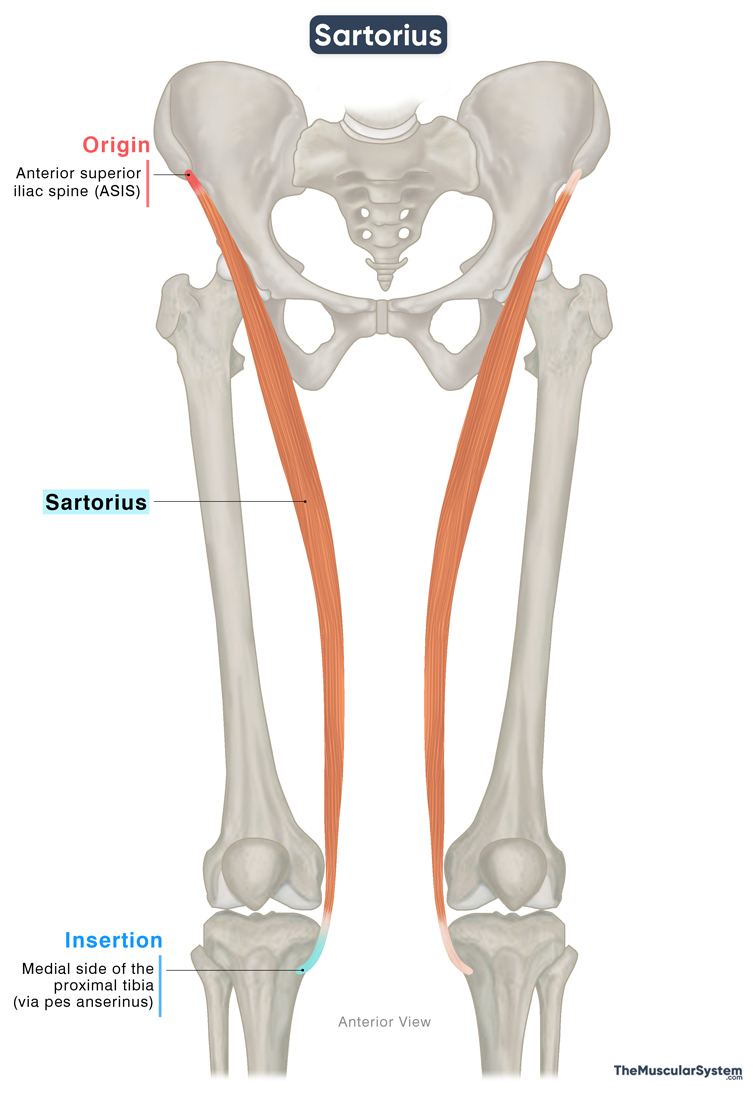
Origin
The muscle originates mainly from the anterior superior iliac spine (ASIS), with some part of its originating tendon arising from the notch just below, between the ASIS and the anterior inferior iliac spine (AIIS).
Insertion
The tendons form a flat, ribbon-like muscle belly and course obliquely and medially downward across the front of the thigh. As it reaches the lateral side of the knee, the muscle fibers narrow into a tendon that curves around the knee joint obliquely to cross the medial condyle of the femur, where it expands into an aponeurotic sheet.
Just posterior to the tendon of the sartorius, the tendons of the semitendinosus and gracilis muscles converge with this aponeurosis to insert into the superior medial surface of the proximal tibia, close to the tibial tuberosity. Here, the tendons of these three muscles form the conjoined tendon called the pes anserinus.
Relations With Surrounding Muscles and Structures
The sartorius is one of the most superficial muscles in the anterior thigh, lying deep to only fascia and skin. The quadriceps femoris muscles (rectus femoris, vastus lateralis, vastus intermedius, and vastus medialis) lie on the deep side of the muscle.
At the proximal end, the muscle originates near the tensor fascia latae, which arises just lateral to its origin. The iliopsoas and adductor longus muscles lie medial to it. At this level, the sartorius forms the lateral boundary of the femoral triangle, a triangular depression in the upper medial thigh that serves as an important anatomical landmark, providing passage for several major nerves and blood vessels. The adductor longus and the inguinal ligament form the medial and superior boundaries of the triangle, respectively.
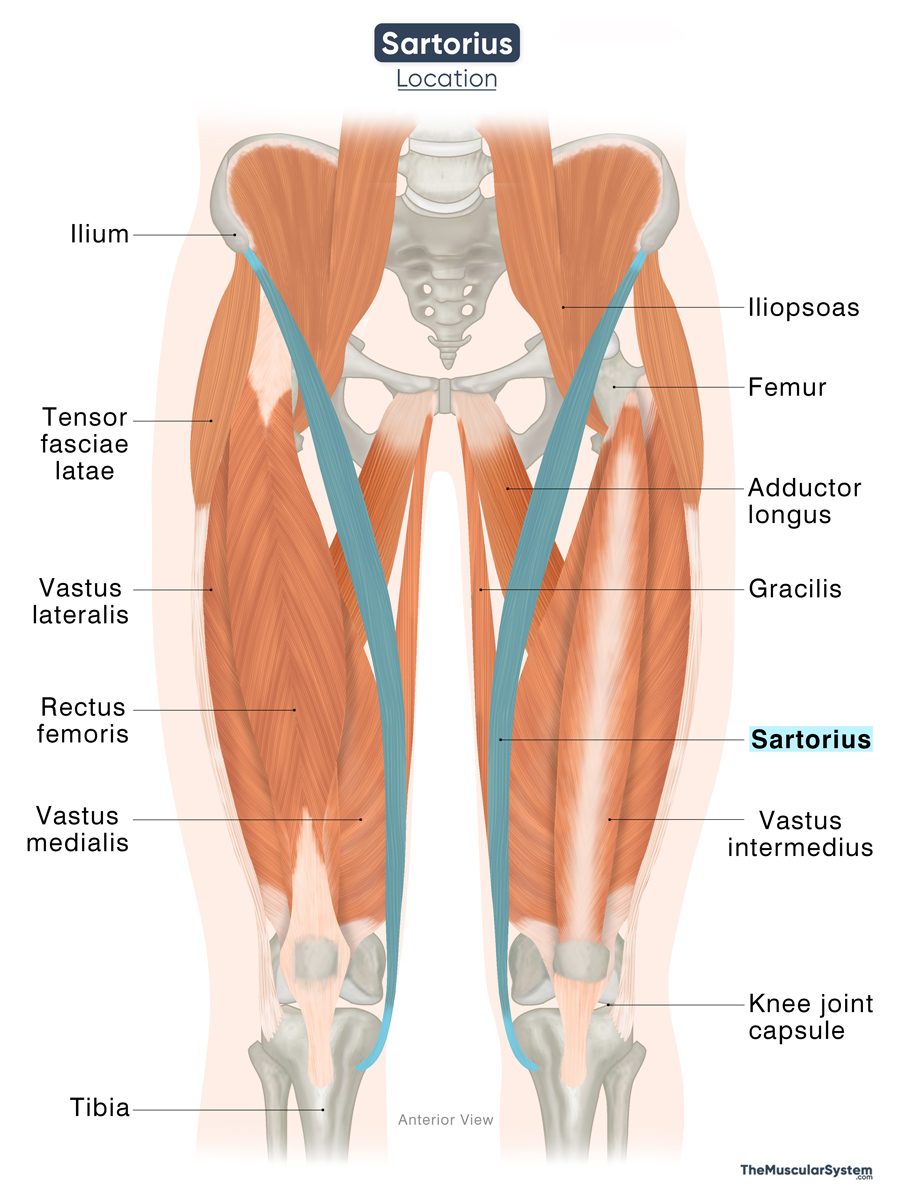
The sartorius also contributes to the roof of the adductor canal, an aponeurotic tunnel in the thigh through which the femoral nerve and vessels pass.
At its distal end, a small bursa lies between the proximal surface of the tibia and the inserting conjoined tendon (pes anserinus) to reduce friction during movement.
Function
| Action | — Flexing, abducting, and laterally rotating the thigh at the hip joint — Flexing and medially rotating the lower leg at the knee joint |
Being a narrow muscle, the sartorius is not capable of producing strong movements on its own. However, due to its attachments both at the hip and knee joints, it is an important synergist muscle that assists several other muscles that move the hip and knee joints.
At the hip joint
The muscle works with the hip flexors (e.g., iliopsoas, rectus femoris), abductors (e.g., gluteus medius, gluteus minimus, tensor fasciae latae), and lateral rotators (e.g., piriformis, the obturators, gemelli, and quadratus femoris), to flex or bend, abduct or move away from the body’s midline, and externally rotate the thigh at the hips.
At the knee joint
Here, when the muscle contracts, it works with the knee flexors (e.g., the hamstrings), and medial rotators (e.g., semimembranosus, semitendinosus, and gracilis) to flex and internally rotate the lower leg at the knee joint.
An action that involves all these movements is when you sit cross-legged.
Antagonists
Due to its synergistic role in several movements, the sartorius does not have a direct antagonist. However, its actions are opposed by different muscle groups depending on the movement.
For its role in flexing the hip, the sartorius is opposed by the hip extensors, such as the gluteus maximus and the hamstrings. Its contribution to hip abduction is countered by the adductor muscles of the thigh. Similarly, its role in external rotation of the hip is opposed by internal rotators like the anterior fibers of the gluteus medius, gluteus minimus, and tensor fasciae latae.
At the knee, where the sartorius assists in flexion, it is opposed by the quadriceps femoris muscles that extend the knee joint.
Innervation
| Nerve | Anterior division of the femoral nerve (L2-L4) |
The muscle receives its innervation from the anterior division of the femoral nerve, rising from the 2nd, 3rd, and 4th lumbar nerve roots (L2, L3, L4).
Blood Supply
| Artery | Femoral artery |
Since the sartorius muscle spans from the hip to the knee, it receives blood supply from multiple sources. The primary vascular supply comes from muscular branches of the femoral artery.
The proximal part of the muscle also receives contributions from the lateral circumflex femoral artery and occasionally from the superficial circumflex iliac artery.
The distal part is additionally supplied by the descending genicular artery, a branch of the femoral artery, along with branches of the medial superior genicular artery from the popliteal artery.
References
- Sartorius: TeachMeAnatomy.info
- Anatomy, Bony Pelvis and Lower Limb: Thigh Sartorius Muscle: NCBI.NLM.NIH.gov
- Sartorius Muscle: Radiopaedia.org
- Sartorius Muscle: Kenhub.com
- Sartorius Muscle: GetBodySmart.com
- Sartorius (Anatomy): PrimaryCareNotebook.com
- Sartorius Muscle: Elsevier.com
Della Barnes, an MS Anatomy graduate, blends medical research with accessible writing, simplifying complex anatomy for a better understanding and appreciation of human anatomy.
- Latest Posts by Della Barnes, MS Anatomy
-
Tensor Tympani
- -
Stapedius
- -
Auricularis Posterior
- All Posts


