

Gracilis
By
Della Barnes, an MS Anatomy graduate, blends medical research with accessible writing, simplifying complex anatomy for a better understanding and appreciation of human anatomy.
Last updated:
08/09/2025Della Barnes, MS Anatomy
UX/UI Designer at - AdobeDella Barnes, an MS Anatomy graduate, blends medical research with accessible writing, simplifying complex anatomy for a better understanding and appreciation of human anatomy.
What is the Gracilis
The gracilis is a long, narrow, unipennate muscle located in the medial compartment of the thigh. It is one of the primary adductor muscles of the hip, alongside the adductor brevis, adductor longus, adductor magnus, and pectineus. The gracilis plays a key role in bringing the thighs together (hip adduction), a movement essential in activities such as horseback riding and skiing.
Anatomy
Location and Attachments
| Origin | The body and inferior ramus of the pubis, and the adjacent part of the ischial ramus |
| Insertion | Medial surface of the tibial shaft on the proximal side (via pes anserinus) |
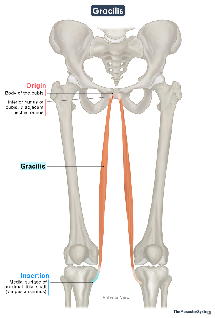
Origin
The muscle originates via a flat aponeurotic band from the anterior body of the pubis, near the inferior aspect of the symphysis pubis. The point of origin extends over the surface of the inferior pubic ramus and partially onto the adjacent ischial ramus.
Insertion
The narrow tendon expands into the muscle belly, which courses vertically downward towards the knee joint. As it descends, the belly tapers and narrows into a long, round tendon that travels posterior to the medial condyle of the femur to reach the proximal tibia. Here, it wraps around the medial tibial condyle, as the round tendon flattens to join the inserting tendons of the sartorius and semitendinosus to form the pes anserinus. Finally, the muscle inserts into the medial surface of the tibial shaft, just below the medial condyle via the pes anserinus.
A few fibers of the tendon may even extend to blend with the deep fascia of the leg.
Relations With Surrounding Muscles and Structures
The proximal side and the muscle belly
The gracilis is the most superficial and medially located muscle in the medial compartment of the thigh. It lies directly beneath the skin and fascia. Medially, it is deep to the fascia lata, while laterally, it is superficial to the adductor brevis and adductor magnus muscles.
The muscle fibers of the gracilis have a spiral orientation as they run down the thigh. So, it is also related to the medial border of the adductor magnus and the inferior border of the adductor brevis. The adductor longus muscle lies laterally to the gracilis.
The distal side and the inserting tendon
At the pes anserinus, the gracilis lies between the tendons of the sartorius muscle lying in front of it, and the semitendinosus lying behind. The tibial collateral ligament, which helps stabilize the knee joint, lies superficial to the gracilis tendon, separated by the anserine bursa, which reduces friction.
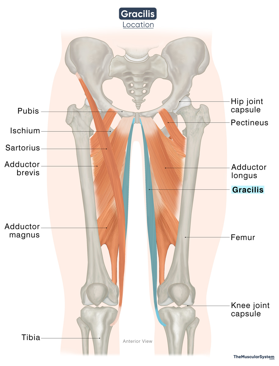
The saphenous nerve and the saphenous branch of the descending genicular artery pass between the tendons of the sartorius and gracilis.
Function
| Action | — Adducting, flexing, and medially rotating the hip joint — Flexing and medially rotating the knee joint |
The gracilis is the only hip adductor that crosses both the hip and knee joints, allowing it to act on both.
At the knee joint
- It is an effective flexor at the knee, working alongside the hamstrings, sartorius, and other knee flexors. It is especially active when the knee is already partially bent, such as during the swing phase of walking, where it assists the hamstrings in controlling leg movement.
- The muscle also contributes to medial rotation of the lower leg at the knee, which means turning the leg inward when the knee is flexed.
A practical example of these combined actions is horseback riding, where the gracilis helps grip the horse with the thighs (hip flexion), maintain knee flexion, and control inward leg rotation for stability and coordination.
At the hip joint (weak)
At the hip, it is the weakest of the adductor muscles. Because of its attachment points at both the hip and knee, the gracilis primarily assists the stronger adductors in drawing the thigh toward the midline. It also contributes, though weakly, to hip flexion and medial rotation. Despite its relatively minor role, the muscle supports movements like squeezing the thighs together during a seated adductor machine workout.
Antagonists
The gracilis does not have a single, direct antagonist muscle. As a hip adductor, it is antagonized by the hip abductors, the gluteus medius, gluteus minimus, and tensor fasciae latae. As a knee flexor, it is opposed by the quadriceps femoris muscles, which extend the knee.
Innervation
| Nerve | Obturator nerve (L2-L3) |
The muscle is innervated by the anterior branch of the obturator nerve, rising from the second and third lumbar spinal nerves (L2-L3). It is a branch of the lumbar plexus.
Blood Supply
| Artery | Medial circumflex femoral artery |
The primary blood supply to the muscle comes from the medial circumflex femoral artery, a branch of the profunda femoris artery. Smaller branches of the profunda femoris may also contribute to its vascular supply.
References
- Anatomy, Bony Pelvis and Lower Limb: Thigh Gracilis Muscle: NCBI.NLM.NIH.gov
- Gracilis Muscle: Elsevier.com
- Gracilis: TeachMeAnatomy.info
- Gracilis Muscle: Kenhub.com
- Gracilis Muscle: IMAIOS.com
- Gracilis Muscle: Radiopaedia.org
Della Barnes, an MS Anatomy graduate, blends medical research with accessible writing, simplifying complex anatomy for a better understanding and appreciation of human anatomy.
- Latest Posts by Della Barnes, MS Anatomy
-
Tensor Tympani
- -
Stapedius
- -
Auricularis Posterior
- All Posts


