

Adductor Brevis
By
Della Barnes, an MS Anatomy graduate, blends medical research with accessible writing, simplifying complex anatomy for a better understanding and appreciation of human anatomy.
Last updated:
22/08/2025Della Barnes, MS Anatomy
UX/UI Designer at - AdobeDella Barnes, an MS Anatomy graduate, blends medical research with accessible writing, simplifying complex anatomy for a better understanding and appreciation of human anatomy.
What is the Adductor Brevis
The adductor brevis is a short, triangular muscle located in the inner thigh region. It belongs to the medial compartment of the thigh, and is one of the five muscles that form the hip adductor group, with the others being the gracilis, pectineus, adductor longus, and adductor magnus.
Apart from its primary role in thigh adduction, the muscle also assists in flexion and rotation, which are essential for movements such as walking and climbing.
Anatomy
Location and Attachments
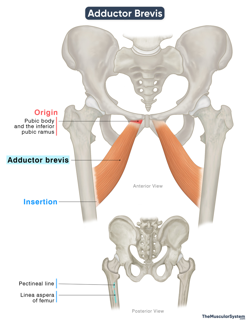
| Origin | The body of the pubis and the inferior pubic ramus |
| Insertion | Medial lip of the linea aspera of the femur, and the pectineal line |
Origin
The muscle originates from a narrow point at the anterior surface of the body of the pubic bone (pubic body) and the lateral side of the inferior pubic ramus.
Insertion
The muscle fibers expand and descend laterally backwards, giving the muscle its characteristic triangular shape. As the fibers approach the femur, they become tendinous and insert via an aponeurosis into the proximal part of the medial lip of the linea aspera, the prominent ridge running along the back of the femoral shaft.
The point of insertion extends to include the pectineal line of the femur, lying just below the lesser trochanter of the femur.
Relations With Surrounding Muscles and Structures
At its origin, the adductor brevis lies between the origin of the gracilis muscle on the medial side and the origin of the obturator externus on the lateral side. In this region, the gracilis and the upper fibers of the adductor magnus lie inferior to the adductor brevis, while the obturator externus lies superior to it.
Further along their muscular bellies, the adductor brevis is positioned posterior or deep to the adductor longus and pectineus muscles, and anterior or superficial to the larger mass of the adductor magnus.
At its insertion, the adductor brevis lies between the insertions of the adductor magnus and the pectineus.
The muscle is also closely related to important neurovascular structures. The medial circumflex femoral artery runs superior to it. The anterior and posterior branches of the obturator nerve course along its anterior and posterior surfaces, respectively. Near its distal attachment to the femur, the second perforating artery pierces the muscle before dividing into ascending and descending branches, which supply the posterior compartment of the thigh.
Adductor Brevis Function
| Action | Adducting the thigh at the hip joint |
The adductor brevis is part of the hip adductor group, and its primary role is to adduct the hip, which means to bring the thigh toward the body’s midline. This action is especially prominent when the thigh is flexed, such as during walking, making the muscle an active participant in the gait cycle.
In addition to adduction, the muscle assists in external rotation of the thigh. It also plays a minor role in hip flexion, helping to bend the thigh at the hip joint. Along with the other muscles in the medial compartment of the thigh, it contributes to stabilizing the pelvis during movement and when standing.
Antagonists
Because its main action is hip adduction, the adductor brevis works in opposition to the hip abductors. These include the gluteus medius, gluteus minimus, and tensor fasciae latae, which pull the thigh away from the midline.
Innervation
| Nerve | Obturator nerve (L2-L4) |
The muscle receives innervation from the anterior branches of the obturator nerve, which arises from the lumbar plexus via the second to fourth lumbar nerve roots (L2–L4).
Blood Supply
| Artery | Medial circumflex femoral artery |
The primary blood supply to the muscle comes from the medial circumflex femoral artery, a major branch of the deep femoral artery. Additional vascular contribution may come from the obturator artery, branching from the internal iliac artery.
References
- Adductor Brevis Muscle: Kenhub.com
- Adductor Brevis: Rad.UW.edu
- Adductor Brevis: Elsevier.com
- Adductor Brevis: TeachMeAnatomy.info
- Adductor Brevis: IMAIOS.com
Della Barnes, an MS Anatomy graduate, blends medical research with accessible writing, simplifying complex anatomy for a better understanding and appreciation of human anatomy.
- Latest Posts by Della Barnes, MS Anatomy
-
Tensor Tympani
- -
Stapedius
- -
Auricularis Posterior
- All Posts


