

Obturator Externus
By
Della Barnes, an MS Anatomy graduate, blends medical research with accessible writing, simplifying complex anatomy for a better understanding and appreciation of human anatomy.
Last updated:
17/07/2025Della Barnes, MS Anatomy
UX/UI Designer at - AdobeDella Barnes, an MS Anatomy graduate, blends medical research with accessible writing, simplifying complex anatomy for a better understanding and appreciation of human anatomy.
What is the Obturator Externus
The obturator externus, or external obturator, is a relatively small, flat, convergent muscle. It is usually classified as part of the medial compartment of the thigh, but some sources group it with the short external rotators of the gluteal region because of its functional association with them. As one of the external rotators of the thigh, it works alongside the obturator internus, piriformis, quadratus femoris, and the gemellus muscles.
Anatomy
Location and Attachments
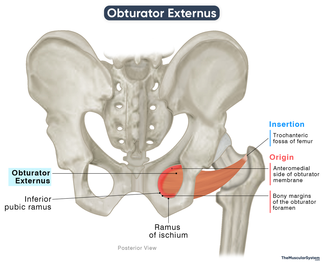
| Origin | The bony margins of the obturator foramen and the anteromedial aspect of the obturator membrane |
| Insertion | Trochanteric fossa of the femur |
Origin
The muscle arises from the bony margins on the medial side of the obturator foramen, specifically from the ramus of the ischium and the inferior pubic ramus. Some muscle fibers also arise from the medial and anterior two-thirds of the obturator membrane and the adjacent tendon.
Insertion
Being a convergent or triangular muscle, the obturator externus has a much broader origin than insertion. From their point of origin, the muscle fibers pass laterally and posteriorly beneath the neck of the femur. They converge into a narrow tendon that courses posterior to the hip joint capsule and inserts into the trochanteric fossa of the femur, located on the medial surface of the greater trochanter.
Relations With Surrounding Muscles and Structures
The muscle sits at the front of the pelvic bone, on its inner surface, covering the obturator foramen, the round opening between the ischium and pubis. It lies deep to the pectineus muscle and the superior portions of the adductor brevis, longus, and magnus muscles. The tendinous part of the obturator externus lies between the quadratus femoris and the neck of the femur, separating the two.
The obturator nerve courses deep to the muscle, with its anterior branch passing from the front of the muscle and the posterior branch piercing it to reach the thigh region.
In some individuals, a bursa, referred to as the obturator externus bursa, may be present between the tendon of the obturator externus and the hip joint capsule. When present, this bursa may help reduce friction at the hip joint during movements.
Function
| Action | Laterally rotating the thigh and helping adduct it when the hip is flexed |
The muscle works in synergy with the other rotators of the thigh and hip region to assist with hip mobility. Here are its main functions:
Rotating the thigh externally: Being a lateral hip rotator, contraction of this muscle, along with the other lateral rotators, helps externally rotate the thigh. An example of this action would be when you pivot or change direction while walking or playing a sport like soccer.
Adducting the thigh: When the hip is in a flexed position, such as in a sitting position or when the knee is raised, the line of pull of the obturator externus shifts slightly. In this position, its contraction can contribute to thigh adduction, drawing the leg back toward the body’s midline.
Stabilizing the hip joint: Since it attaches to the ischium, pubis, and the femur near the greater trochanter, the obturator externus plays a vital role in stabilizing the femoral head within the acetabulum.
Antagonists
The obturator externus muscle does not have any exclusive antagonists. However, the medial rotators of the thigh, like the gluteus medius and minimus and the tensor fascia latae, work antagonistically to the lateral hip rotators.
Additionally, when the hip is flexed, the slight adduction assisted by the obturator externus is opposed by the abductors of the thigh, including the obturator internus, gluteus medius, and gluteus minimus. However, these are not the primary antagonists to the muscle.
Innervation
| Nerve | Posterior branch of the obturator nerve (L3-L4) |
The posterior branch of the obturator nerve gives off a narrow branch to innervate the obturator externus as it pierces the muscle on its way to the thigh. The fibers supplying the obturator externus arise primarily from the third and fourth lumbar nerves (L3–L4).
Blood Supply
| Artery | Obturator and medial circumflex femoral arteries |
Blood supply to this muscle comes from two sources. The anterior branch of the obturator artery, which itself is a branch of the internal iliac artery, supplies the muscle. Another source of blood supply is the medial circumflex femoral artery, which branches from the profunda femoris artery, the largest deep branch of the femoral artery (the primary arterial supply to the lower limbs).
Anatomical variation may exist, with the muscle receiving blood from either one or both of these arteries.
References
- Anatomy, Abdomen and Pelvis, Obturator Muscles: NCBI.NLM.NIH.gov
- Obturator Externus: Elsevier.com
- Obturator Externus: IMAIOS.com
- Obturator Externus: TeachMeAnatomy.info
- Obturator Externus: Rad.Washington.edu
- Obturator Externus Muscle: Kenhub.com
Della Barnes, an MS Anatomy graduate, blends medical research with accessible writing, simplifying complex anatomy for a better understanding and appreciation of human anatomy.
- Latest Posts by Della Barnes, MS Anatomy
-
Tensor Tympani
- -
Stapedius
- -
Auricularis Posterior
- All Posts


