

Piriformis
By
Della Barnes, an MS Anatomy graduate, blends medical research with accessible writing, simplifying complex anatomy for a better understanding and appreciation of human anatomy.
Last updated:
14/08/2025Della Barnes, MS Anatomy
UX/UI Designer at - AdobeDella Barnes, an MS Anatomy graduate, blends medical research with accessible writing, simplifying complex anatomy for a better understanding and appreciation of human anatomy.
What is the Piriformis
The piriformis is a small convergent muscle in the buttocks or gluteal region. The Latin word “piriformis” means “pear-shaped,” which refers to the shape of the muscle. It belongs to the group of lateral rotators of the hip along with the quadratus femoris, the gemellus, and the obturator muscles. The muscle plays a crucial role in rotating and moving the thigh in various directions.
Anatomy
Location and Attachments
| Origin | Anterior surface of the sacrum (S2-S4) |
| Insertion | Greater trochanter of femur |
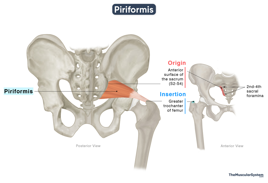
Origin
The muscle originates from the anterior surface, and partially the lateral surface, of the sacrum within the pelvic region. Its fibers arise in three slips from the regions between the second and fourth sacral foramina (S2-S4). Some part of the muscle also originates from the superior part of the gluteal surface of the ilium, the upper border of the greater sciatic notch, and the sacrotuberous ligament.
Insertion
From its origin, the muscle belly courses laterally and inferiorly, passing through the greater sciatic foramen to exit the bony pelvis and enter the gluteal region. The muscle fibers then converge into a slender tendon, which inserts onto the top of the greater trochanter of the femur. The insertion tendon may occasionally fuse with the conjoined tendon of the superior and inferior gemelli and the obturator internus, which share a common insertion point.
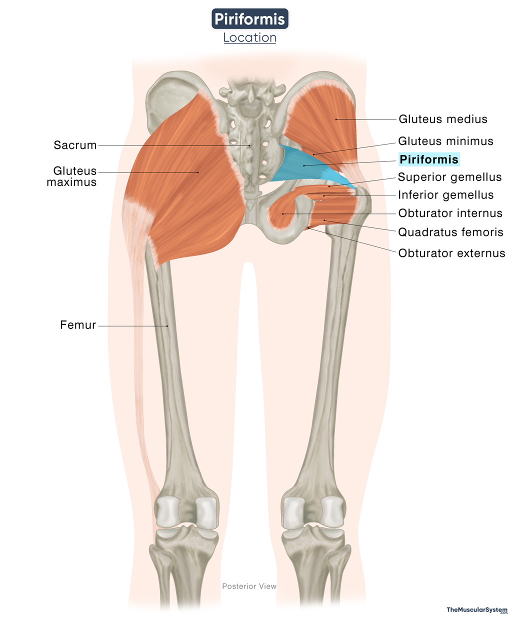
Piriformis Relations With Surrounding Muscles and Structures
The piriformis muscle is situated in the deep gluteal region, where it lies deep to the gluteus maximus and inferior to the gluteus medius, which runs parallel and superior to it. The gluteus maximus also covers the posterior aspect of the muscle. The coccygeus and superior gemellus muscles lie inferomedial and inferior to the piriformis, respectively. In front of the piriformis lies the hip joint capsule and the sacral plexus, the network of nerves of the pelvic region.
As it passes through the greater sciatic foramen, the piriformis occupies a substantial portion of the greater sciatic notch of the ilium. In doing so, it divides the foramen into two compartments: the suprapiriform space lying above the muscle, and the infrapiriform space lying below the muscle. It makes the muscle an important reference point for both neural and vascular structures traversing this area.
The superior gluteal nerve and vessels typically pass superior to the muscle, while the inferior gluteal nerve and vessels pass below it. The sciatic nerve, the longest nerve in the human body, courses deep to the muscle in about 80% of people. However, anatomical variations do exist where the nerve may pass through or even above the muscle.
Due to its close anatomical relationship with the sciatic nerve, the piriformis muscle may sometimes compress or irritate the nerve, leading to pain, tingling, or numbness along the back of the thigh and leg (sciatica). This condition is known as piriformis syndrome.
Function
| Action | Laterally rotating the thigh, helping abduct the hip from a flexed position, and stabilizing the hip |
The muscle plays a vital role in maintaining flexibility of the hip and thigh region.
Rotating the thigh externally: The piriformis works alongside other lateral hip rotators to help externally rotate the thigh. This movement is vital in activities like changing direction while walking or pivoting during sports such as soccer.
Abducting the thigh: When the hip is flexed to 90°, the piriformis assists the gluteus medius and minimus in abducting the thigh. Which means it helps move the thigh outward, away from the body. A good example of this is swinging your leg to the side while sitting in a chair.
Stabilizing the hip joint: Having its attachment to the greater trochanter of the femur, the piriformis helps stabilize the femoral head within the acetabulum, the cup-shaped socket of the pelvis that forms the hip joint.
Antagonists
The gluteus medius and gluteus minimus assist in medially rotating the thigh at the hip joint, which is the opposite action of the lateral rotators. In this sense, they can be considered antagonists to the piriformis, which acts as a lateral rotator of the hip.
Innervation
| Nerve | Piriformis nerve (S1-S2) |
The muscle gets innervated by the piriformis nerve, also called the nerve to piriformis, rising from the anterior rami of the 1st and 2nd sacral nerves (S1-S2).
Blood Supply
| Artery | Inferior and superior gluteal, internal pudendal, and lateral sacral arteries |
Blood supply to the muscle comes from several branches of the internal iliac artery, including the Inferior and superior gluteal arteries, the internal pudendal artery, and the lateral sacral artery.
References
- Anatomy, Bony Pelvis and Lower Limb: Piriformis Muscle: NCBI.NLM.NIH.gov
- Piriformis: Healthline.com
- Piriformis: Rad.Washington.edu
- Piriformis Muscle: Elsevier.com
- Piriformis Muscle: Definition, Location & Function: Study.com
- Piriformis Muscle: Kenhub.com
- Piriformis Muscle: Radiopaedia.org
- What is the Piriformis Muscle? A Guide to Understanding Function, Impact, and Remedies: PrincetonOrthopaedic.com
- Piriformis Muscle: IMAIOS.com
Della Barnes, an MS Anatomy graduate, blends medical research with accessible writing, simplifying complex anatomy for a better understanding and appreciation of human anatomy.
- Latest Posts by Della Barnes, MS Anatomy
-
Tensor Tympani
- -
Stapedius
- -
Auricularis Posterior
- All Posts


