

Lateral Rotators of the Hip
By
Della Barnes, an MS Anatomy graduate, blends medical research with accessible writing, simplifying complex anatomy for a better understanding and appreciation of human anatomy.
Last updated:
29/08/2025Della Barnes, MS Anatomy
UX/UI Designer at - AdobeDella Barnes, an MS Anatomy graduate, blends medical research with accessible writing, simplifying complex anatomy for a better understanding and appreciation of human anatomy.
What are the Lateral Rotators of the Hip
The lateral rotators of the hip are a group of six short muscles located deep in the gluteal region of the hip. Their main function is to rotate the thigh laterally, or outward at the hip joint. This action is vital in maintaining balance, stability, and changing directions during walking, running, or other activities.
List of Muscles in the Lateral Rotator Group With Their Anatomy
Apart from their common general location, the muscles that laterally rotate the hip also share the following characteristics:
- All these muscles originate from the hip bone.
- They all insert into the femur, specifically into its proximal extremity.
- All of them are innervated by branches of the sacral plexus (L4–S2), except the obturator externus, which is innervated by the lumbar plexus (L1-L4).
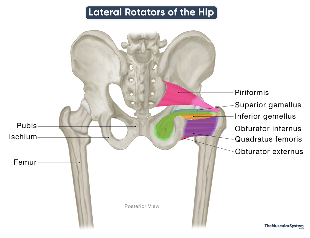
Here’s a list of these muscles, arranged from superficial to deep:
| Muscle | Origin | Insertion | Innervation | Blood Supply |
|---|---|---|---|---|
| Piriformis | Anterior surface of sacrum (S2–S4) | Greater trochanter of femur | Nerve to piriformis (S1–S2) | Inferior and superior gluteal arteries, internal pudendal artery, lateral sacral arteries |
| Gemelli muscles | ||||
| – Superior gemellus | Ischial spine | Trochanteric fossa of femur (via tendon of obturator internus) | Nerve to obturator internus (L5–S2) | Inferior gluteal artery |
| – Inferior gemellus | Ischial tuberosity (upper posterior surface) | Trochanteric fossa of femur (via tendon of obturator internus) | Nerve to quadratus femoris (L5–S1) | Inferior gluteal artery |
| Obturator internus* | — Ischiopubic ramus— Posterior surface of obturator membrane | Trochanteric fossa of femur | Nerve to obturator internus (L5–S2) | Inferior gluteal artery |
| Quadratus femoris | Lateral surface of ischial tuberosity | Quadrate tubercle of femur | Nerve to quadratus femoris (L4–S1) | Inferior gluteal artery, medial circumflex femoral artery |
| Obturator externus | — Margins of the obturator foramen— Anteromedial surface of obturator membrane | Trochanteric fossa of femur | Posterior branch of obturator nerve (L3–L4) | Obturator artery, medial circumflex femoral artery |
Apart from laterally rotating the thigh at the hip, many of these muscles also assist in adduction when the hip is flexed, effectively bringing the thighs closer together. A common example of this action is crossing the legs while sitting in a chair.
Beyond these primary lateral rotators, other muscles such as the gluteus maximus, sartorius, iliopsoas, and the hip adductors can also contribute to lateral rotation of the hip, particularly when the joint is in extension or partial flexion.
On the other hand, the medial rotators of the hip, the gluteus medius, gluteus minimus, tensor fasciae latae, and the adductor brevis, longus, and magnus, act as antagonists to this movement, producing the opposite movement by rotating the thigh medially at the hip joint.
References
- Learn Muscle Anatomy: Lateral Rotators: VisibleBody.com
- Muscles of the Gluteal Region: TeachMeAnatomy.info
Della Barnes, an MS Anatomy graduate, blends medical research with accessible writing, simplifying complex anatomy for a better understanding and appreciation of human anatomy.
- Latest Posts by Della Barnes, MS Anatomy
-
Tensor Tympani
- -
Stapedius
- -
Auricularis Posterior
- All Posts


