

Hip Adductors
By
Della Barnes, an MS Anatomy graduate, blends medical research with accessible writing, simplifying complex anatomy for a better understanding and appreciation of human anatomy.
Last updated:
22/08/2025Della Barnes, MS Anatomy
UX/UI Designer at - AdobeDella Barnes, an MS Anatomy graduate, blends medical research with accessible writing, simplifying complex anatomy for a better understanding and appreciation of human anatomy.
What Are the Hip Adductors
The hip adductors are a group of five muscles in the lower limb that have the primary function of adducting the thigh at the hip joint. This action is crucial for various everyday activities, from walking, running, and climbing, to dancing, horseback riding, and other sports.
Though the group is called the “hip” adductors based on the joint they act on, the muscles in this group belong to the medial compartment of the thigh.
List of Hip Adductor Muscles With Their Anatomy
The muscles that adduct the hip share these characteristics:
- All of them originate from the hip bone, specifically the pubis and ischium.
- All insert into the femur, except the gracilis, which inserts into the tibia.
- All receive innervation from the obturator nerve, a branch of the lumbar plexus.
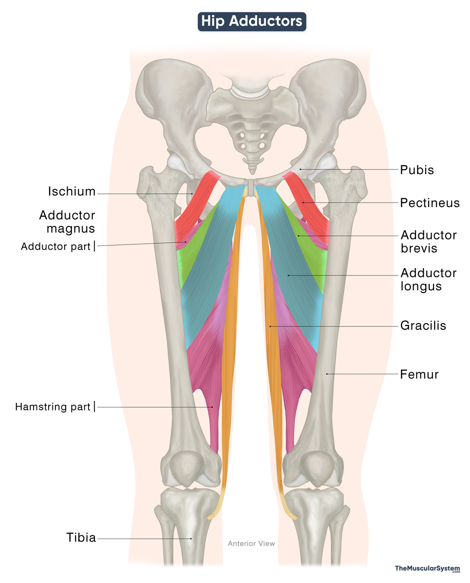
From superficial to deep, these muscles are:
| Muscle | Origin | Insertion | Innervation | Blood Supply |
|---|---|---|---|---|
| Gracilis | — Body and inferior ramus of the pubis — Adjacent ischial ramus | Medial surface of proximal tibia (via pes anserinus) | Obturator nerve (anterior branch, L2–L3) | Medial circumflex femoral artery |
| Pectineus | Pectineal line of the pubis | Pectineal line of the femur | Femoral nerve (L2–L3); additional supply from the obturator nerve (anterior branch, L2–L3) | Medial circumflex femoral artery (superficial portion of the muscle), obturator artery (deep portion) |
| Adductor Longus | Anterior surface of the body of the pubis | Middle third of the linea aspera of the femur | Obturator nerve (anterior branch, L2–L4) | Profunda femoris artery, obturator artery, medial circumflex femoral artery (proximal portion of the muscle) |
| Adductor Brevis | Body and inferior ramus of the pubis | Pectineal line and proximal linea aspera | Obturator nerve (anterior branch, L2–L4) | Medial circumflex femoral artery |
| Adductor Magnus | ||||
| — Adductor Part | — Inferior pubic ramu — Ischial ramus | — Gluteal tuberosity of the femur — Linea aspera — Medial supracondylar line of the femur | — Obturator nerve (posterior branch, L2–L4) | Perforating branches of profunda femoris artery, and medial circumflex femoral artery (proximal portion of the muscle) |
| — Hamstring Part | — Ischial tuberosity | — Medial supracondylar line — Adductor tubercle of the femur | — Tibial nerve (L4) | Same as the adductor part |
Note: The “hamstring part” of the adductor magnus differs from the rest of the adductors because it is innervated by the tibial branch of the sciatic nerve, like the hamstring muscles. This is why it’s called the hamstring part. But since it inserts on the femur and does not cross the knee, it is not considered a true hamstring muscle.
What Do the Hip Adductors Do
Primary Functions
The hip adductors work mainly to bring the thigh inward toward the midline of the body, a movement known as adduction.
- When only one leg is working, these muscles are strongest if the hip begins in the normal standing position. An example of this movement would be when you cross your legs while standing.
- When both legs are working together, the adductors are strongest during movements that also involve flexing or bending, and extending the hips and knees, such as weight-bearing exercises or horseback riding.
Secondary Functions
The muscles also help with the following movements at the hip joint:
- Flexing or bending the thigh, like when you climb stairs.
- Extending, or straightening the thigh, like pushing the leg back during walking.
- Medially rotating or turning the thigh inward, like when changing direction while walking.
- Laterally rotating or turning the thigh outward, like crossing one ankle over the opposite knee while sitting.
Antagonists
The hip abductors, gluteus medius, gluteus minimus, and tensor fasciae latae, act as the primary antagonists of the adductors, producing abduction by moving the thigh away from the midline.
During dynamic activities such as walking, running, or balancing on one leg, the abductors and adductors work in opposition yet in coordination to maintain pelvic stability and control the range of hip movement.
References
- Hip Adductors: Kenhub.com
- Muscles in the Medial Compartment of the Thigh: TeachMeAnatomy.info
- Hip Adductors: Sciencedirect.com
Della Barnes, an MS Anatomy graduate, blends medical research with accessible writing, simplifying complex anatomy for a better understanding and appreciation of human anatomy.
- Latest Posts by Della Barnes, MS Anatomy
-
Tensor Tympani
- -
Stapedius
- -
Auricularis Posterior
- All Posts


