

Adductor Hallucis
By
Della Barnes, an MS Anatomy graduate, blends medical research with accessible writing, simplifying complex anatomy for a better understanding and appreciation of human anatomy.
Last updated:
29/10/2025Della Barnes, MS Anatomy
UX/UI Designer at - AdobeDella Barnes, an MS Anatomy graduate, blends medical research with accessible writing, simplifying complex anatomy for a better understanding and appreciation of human anatomy.
What is the Adductor Hallucis
The adductor hallucis is a two-headed, small, intrinsic muscle located at the middle of the sole of the foot. It is one of the three muscles in the third layer of plantar muscles, with the other two being the flexor hallucis brevis and flexor digiti minimi brevis.
As implied by its name, it is the primary muscle responsible for adduction of the big toe, an action that helps with movements like walking and running.
Anatomy
The muscle has one oblique head and one transverse head, with different origins, but the same insertion.
Location and Attachments
| Origin | — Oblique head: The 2nd-4th metatarsal base — Transverse head: Plantar metatarsophalangeal ligaments of the 3rd-5th toes |
| Insertion | The lateral side of the proximal phalangeal base of the big toe |
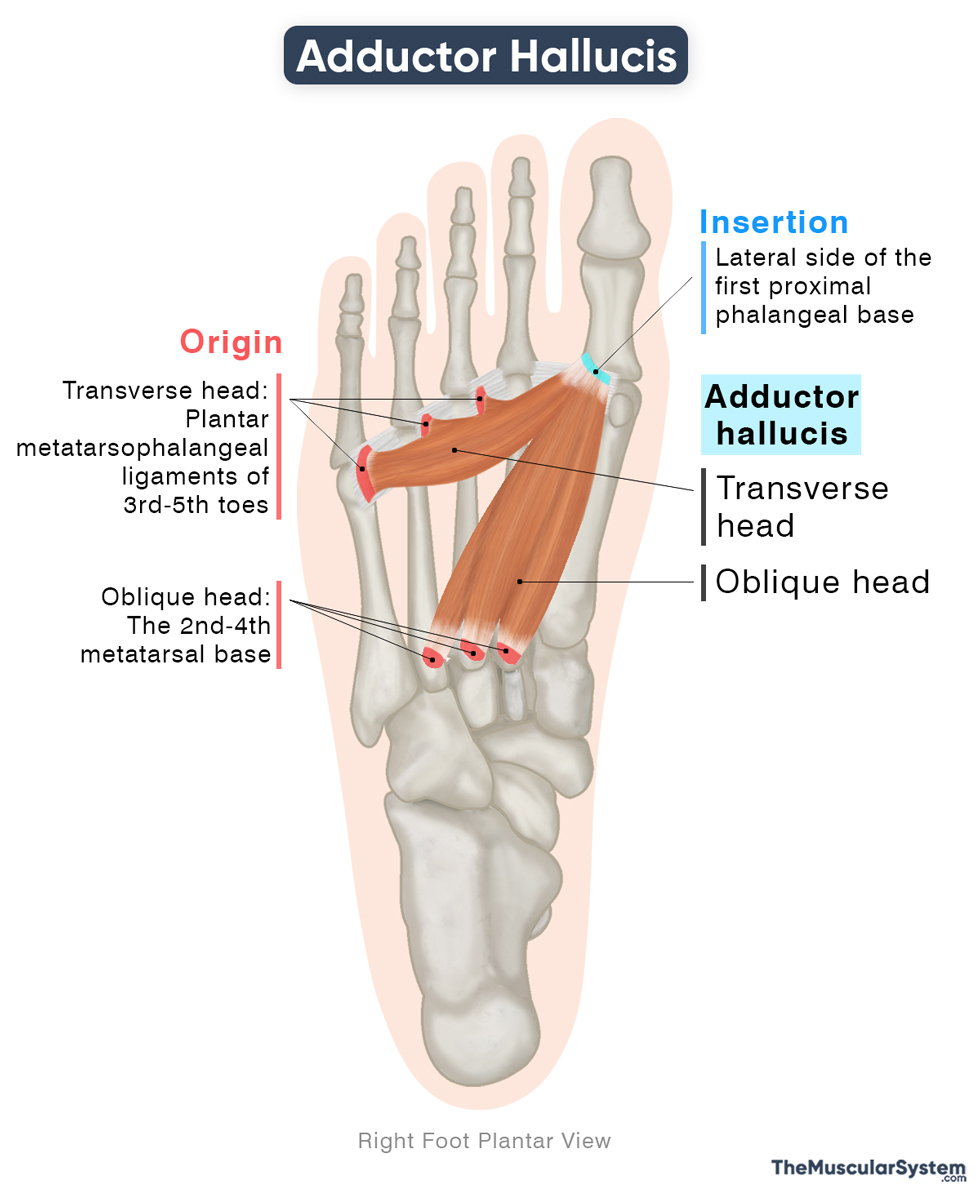
Adductor Hallucis Origin
Oblique Head
The oblique head is the thicker and more muscular portion of the adductor hallucis, occupying the space beneath the second, third, and fourth metatarsal bones. It originates as muscular slips from the bases of the second to fourth metatarsals. Additionally, some fibers may arise from the fibrous sheath surrounding the tendon of the peroneus longus.
Transverse head
The transverse head consists of slender, flat muscle bands located beneath the second to fourth toes. It originates as three muscular slips from the plantar metatarsophalangeal ligaments of the third to fifth toes, as well as from the deep transverse metatarsal ligaments that connect these joints.
Variations may occur where the transverse head rises only from the third and fourth toes.
Adductor Hallucis Insertion
The fibers of the oblique head unite to form a fleshy belly that runs forward, diagonally, and medially, toward the big toe, where it joins the lateral head of the flexor hallucis brevis.
Similarly, the muscular bands of the transverse head merge medially to converge with the oblique head.
Together, both heads insert into the lateral side of the base of the proximal phalanx of the big toe.
Relations With Surrounding Muscles and Structures
- Inferiorly, it is related to the first and second dorsal and plantar interossei muscles, which belong to the fourth layer of plantar muscles.
- Superiorly, it lies deep to the lumbrical muscles of the second layer, the tendon of the flexor digitorum brevis from the first layer, and the tendon of the flexor digitorum longus originating from the leg.
- Medially and proximally, it is in relation to the flexor hallucis brevis, which belongs to the third layer of plantar muscles.
The muscle is also related to several nerves and blood vessels. Across its dorsal surface, the deep plantar arterial arch and the deep branch of the lateral plantar nerve pass over the oblique head, while the plantar metatarsal arteries pass over the transverse head. The proper plantar digital nerves pass underneath both heads.
Function
| Action | Adduction of the big toe at the metatarsophalangeal joint |
Role in moving the big toe
- When it contracts, the muscle adducts the big toe at the metatarsophalangeal joint, pulling it toward the second toe.
- It also assists the flexor hallucis longus in flexing the big toe at the same joint, bending it downward toward the sole.
Through these actions, the muscle plays a vital role in the gait cycle by stabilizing the front of the foot as the heel lifts during walking, thereby aiding smooth forward movement.
Role in stabilizing the foot
The muscle also contributes to the stabilization of the foot’s arches, particularly the transverse arch, which is reinforced by the horizontally oriented transverse head.
Antagonists
The abductor hallucis acts as the primary antagonist of this muscle, as it is responsible for abducting the big toe, moving it away from the second toe, which is the opposite of adduction.
Innervation
| Nerve | Lateral plantar nerve (S2-S3) |
Both heads of the muscle receive innervation from the deep branch of the lateral plantar nerve, carrying branches from the second and third sacral nerve roots (S2-S3). The lateral plantar nerve is one of the two terminal branches of the tibial nerve.
Blood Supply
| Artery | Medial and lateral plantar arteries |
The primary blood supply to this muscle comes from the medial and lateral plantar arteries, both of which branch from the posterior tibial artery. The plantar arch and its branches, the first to fourth plantar metatarsal arteries, provide additional blood supply.
References
- Adductor Hallucis (Left): Elsevier.com
- Adductor Hallucis: TeachMeAnatomy.info
- Adductor Hallucis Muscle: Kenhub.com
- Adductor Hallucis: IMAIOS.com
- Adductor Hallucis Muscle: Radiopaedia.org
Della Barnes, an MS Anatomy graduate, blends medical research with accessible writing, simplifying complex anatomy for a better understanding and appreciation of human anatomy.
- Latest Posts by Della Barnes, MS Anatomy
-
Tensor Tympani
- -
Stapedius
- -
Auricularis Posterior
- All Posts


