

Flexor Digitorum Brevis
By
Della Barnes, an MS Anatomy graduate, blends medical research with accessible writing, simplifying complex anatomy for a better understanding and appreciation of human anatomy.
Last updated:
17/10/2025Della Barnes, MS Anatomy
UX/UI Designer at - AdobeDella Barnes, an MS Anatomy graduate, blends medical research with accessible writing, simplifying complex anatomy for a better understanding and appreciation of human anatomy.
What is the Flexor Digitorum Brevis
The flexor digitorum brevis (FDB) is a broad, fusiform muscle located in the central region of the plantar surface or sole of the foot. It belongs to the superficial, or first, layer of the plantar muscles, together with the abductor hallucis and abductor digiti minimi.
Functionally, the FDB flexes the lateral four toes and contributes to maintaining foot stability during standing and walking.
Anatomy
Location and Attachments
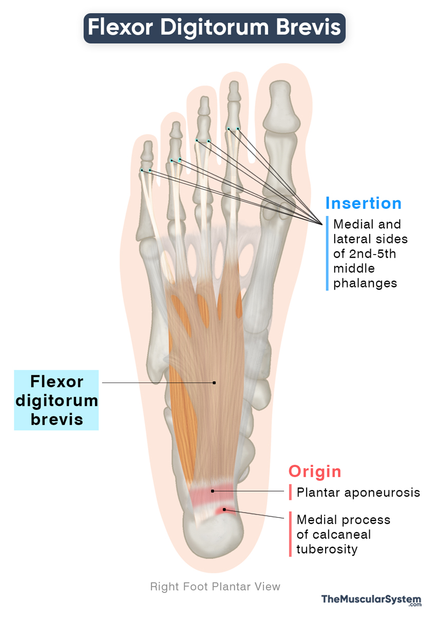
| Origin | Medial process of the calcaneal tuberosity, the plantar aponeurosis, and the intermuscular septum of the foot |
| Insertion | Medial and lateral surfaces of the 2nd to 5th middle phalanges |
Origin
Like the other two muscles in the first layer of the plantar muscles, the flexor digitorum brevis originates from the medial process/tubercle of the calcaneal tuberosity. Additional fibers arise from the central part of the plantar aponeurosis and the intermuscular septa separating it from adjacent muscles.
Insertion
The FDB has a large muscle belly that occupies the central portion of the plantar surface of the foot. It courses forward, narrowing and dividing into four tendinous slips about halfway across the sole. Each of these slips corresponds to the second through fifth toes, running along the plantar surfaces of their respective metatarsals.
At the base of the proximal phalanx of each of these toes, the FDB tendons divide to form a tendinous tunnel to allow passage to the tendons of the flexor digitorum longus to pass through on their way to the distal phalanges. The two portions of each FDB tendon reunite and continue forward before splitting completely into two slips for insertion on the medial and lateral sides of the middle phalanx of the second to fifth toes.
Variation
The muscle often shows structural variations, especially in the slip to the little toe, which may be narrower than the others or even absent in some people. When the slip is missing, its function may be taken over by a small muscle originating from either the flexor digitorum longus tendon or the quadratus plantae muscle.
Relations With Surrounding Muscles and Structures
Within the plantar surface, it lies lateral to the abductor hallucis and medial to the abductor digiti minimi. Superficially, it is covered by the plantar aponeurosis, a dense connective tissue layer that forms the sole. Deep to the FDB are the quadratus plantae and lumbrical muscles, along with the tendons of the flexor digitorum longus.
Its tendons pass medial to the common plantar digital nerves and vessels, while the muscle itself lies superficial to the lateral plantar nerve and vessels.
Function
| Action | Flexion of the 2nd to 5th digits |
It primarily acts to flex the four lateral toes at both their metatarsophalangeal and proximal interphalangeal joints. Unlike the flexor digitorum longus, which initiates flexion at the distal interphalangeal joints, this muscle focuses on the more proximal joints of the toes. Together, these two muscles work in synergy to maintain stability during the gait cycle, ensuring the toes remain in firm contact with the ground.
Additionally, the FDB contributes to the formation of the longitudinal arch of the foot, helping with balance and stability during walking and running.
This muscle is analogous to the flexor digitorum superficialis of the hand. Each serves as an intrinsic flexor for the lateral four digits, acting on toes 2-5 in the foot and fingers 2-5 in the hand, facilitating flexion and stability.
Antagonists
The extensor digitorum longus and extensor digitorum brevis are the primary antagonists of this muscle, as they extend the toes, which is the opposite action of toe flexion.
Innervation
| Nerve | Medial plantar nerve (S1-S3) |
The medial plantar nerve, the larger of the two terminal branches of the tibial nerve, innervates this muscle. It originates from the first to third sacral nerve roots (S1–S3) of the sacral plexus.
Blood Supply
| Artery | Medial plantar, lateral plantar, plantar metatarsal, and common plantar digital arteries |
It receives its primary blood supply from the medial and lateral plantar arteries, both branches of the posterior tibial artery. Additional supply is provided by the plantar metatarsal arteries and their branches, the common plantar digital arteries, arising from the plantar arch.
References
- Flexor Digitorum Brevis: Elsevier.com
- Flexor Digitorum Brevis: TeachMeAnatomy.info
- Flexor Digitorum Brevis Muscle: Kenhub.com
- Flexor Digitorum Brevis: HealthLine.com
- Flexor Digitorum Brevis Muscle: Radiopaedia.org
- Some Variations of the Musculus Flexor Digitorum Brevis: PubMed.NCBI.NLM.NIH.gov
Della Barnes, an MS Anatomy graduate, blends medical research with accessible writing, simplifying complex anatomy for a better understanding and appreciation of human anatomy.
- Latest Posts by Della Barnes, MS Anatomy
-
Tensor Tympani
- -
Stapedius
- -
Auricularis Posterior
- All Posts


