

Palatopharyngeus
By
Della Barnes, an MS Anatomy graduate, blends medical research with accessible writing, simplifying complex anatomy for a better understanding and appreciation of human anatomy.
Last updated:
26/12/2025Della Barnes, MS Anatomy
UX/UI Designer at - AdobeDella Barnes, an MS Anatomy graduate, blends medical research with accessible writing, simplifying complex anatomy for a better understanding and appreciation of human anatomy.
What is the Palatopharyngeus
The palatopharyngeus, also known as the palatopharyngeal or pharyngopalatinus muscle, is a long, narrow, paired skeletal muscle that extends from the soft palate to the upper part of the pharynx. Due to this reason, it is classified as a muscle of the head with the other soft palate muscles, as well as a longitudinal pharyngeal muscle, with the stylopharyngeus and salpingopharyngeus.
When you open your mouth wide, the muscle can be seen as the posterior pillar of the arch at the back of the mouth and the beginning of the throat. It works with the longitudinal pharyngeal muscles to elevate the pharynx and help with swallowing.
Anatomy
Location and Attachments
| Origin | Posterior margin of the hard palate and the palatine aponeurosis |
| Insertion | Back of the thyroid cartilage and the pharyngeal constrictor muscles |
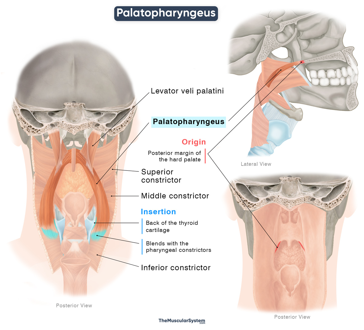
Origin
The Palatopharyngeus muscle originates from the posterior margin of the hard palate, at the back of the oral cavity. Part of the muscle also originates from the palatine aponeurosis, the tough, fibrous sheet that lends shape and structure to the soft palate. At the origin, the levator veli palatini divides the muscle fibers into two parts:
- The anterior fascicles: The thicker part of the muscle, located in the soft palate, it runs between the levator veli palatini and the tensor veli palatini muscles. It joins the fibers from the opposite side at the center.
- The posterior fascicles: The narrower part lies in the oral mucosa, and like the anterior part, merges with the contralateral fibers at the center.
The two parts merge to form the muscle belly at the posterolateral margin of the soft palate. The muscle belly is narrower than the originating and inserting parts.
Insertion
The fibers course laterally and posteriorly downward along the wall of the pharynx. They gradually spread out, inserting into the back of the thyroid cartilage and merging with the pharyngeal constrictors, forming a fan-shaped insertion. A few of the fibers may course to the midline and blend with the fibers from the contralateral palatopharyngeus.
Relations With Surrounding Muscles and Structures
With other muscles and structures near the soft palate
At its origin near the soft palate, the muscle is closely related to other soft-palate structures. As already mentioned, the levator veli palatini divides the muscle into the anterior and posterior fascicles. The musculus uvulae, the small paired muscle that helps control the uvula, lies medial to it.
It lies immediately deep to the pharyngeal mucosa. The muscle forms the posterior pillar of the fauces, the opening of the oropharynx right behind the oral cavity. The palatoglossus, a key muscle in the soft palate, forms the anterior pillar of the fauces, with the two muscles being separated by the tonsillar fossa, which contains the palatine tonsil.
With the longitudinal muscles of the pharynx
Inferiorly, where the two fascicles of the muscle reunite, the salpingopharyngeus muscle blends with the palatopharyngeus. As it descends further, it lies adjacent to the stylopharyngeus muscle anteromedially. Both muscles course inferiorly toward their insertion along the posterior border of the thyroid cartilage.
Function
| Action | Helps with swallowing by elevating the pharynx, and closing off the entrance to the nasopharynx |
During swallowing, the soft palate is raised and stretched by the muscles levator veli palatini and tensor veli palatini. Once the chewed food or bolus reaches the back of the throat, the palatopharyngeus contracts to raise and shorten the pharynx. This pulls the palatopharyngeal arches closer together, narrowing the pharyngeal lumen. It forms a transient bulge or prominence in the posterior pharyngeal wall (Passavant’s ridge), with contribution from the fibers of the superior pharyngeal constrictor. This ridge temporarily closes off the nasopharynx, preventing the bolus from entering it.
Finally, the oblique orientation of the palatopharyngeus fibers helps guide and propel the bolus downward through the lower pharynx toward the esophagus.
Antagonists
The palatopharyngeus has no single, direct antagonist as its actions are part of a coordinated sequence. While the palatopharyngeus depresses the soft palate and narrows the pharynx during swallowing, muscles that elevate and stabilize the soft palate, especially the levator veli palatini, perform the opposite action. However, these muscles act in sequence rather than in strict opposition.
Innervation
| Nerve | Pharyngeal plexus (CN X) |
It receives innervation from the same source as the other pharyngeal muscles, via branches of the pharyngeal plexus, which itself arises from the vagus nerve (CN X).
Blood Supply
| Artery | Ascending palatine artery, greater palatine artery, and the ascending pharyngeal artery, via its pharyngeal branch |
Blood supply to the muscle comes from the ascending palatine artery, which is a branch of the facial artery, the greater palatine artery, a branch of the maxillary artery, and the pharyngeal trunk of the ascending pharyngeal artery.
References
- Palatopharyngeus Muscle: Kenhub.com
- Palatopharyngeus: TeachMeAnatomy.info
- Palatopharyngeus Muscle: Elsevier.com
- Palatopharyngeus Muscle: Radiopaedia.org
- Palatopharyngeus: Meddean.LUC.edu
Della Barnes, an MS Anatomy graduate, blends medical research with accessible writing, simplifying complex anatomy for a better understanding and appreciation of human anatomy.
- Latest Posts by Della Barnes, MS Anatomy
-
Tensor Tympani
- -
Stapedius
- -
Auricularis Posterior
- All Posts


