

Stapedius
By
Della Barnes, an MS Anatomy graduate, blends medical research with accessible writing, simplifying complex anatomy for a better understanding and appreciation of human anatomy.
Last updated:
12/02/2026Della Barnes, MS Anatomy
UX/UI Designer at - AdobeDella Barnes, an MS Anatomy graduate, blends medical research with accessible writing, simplifying complex anatomy for a better understanding and appreciation of human anatomy.
The stapedius is the smallest skeletal muscle in the human body and one of the two inner ear muscles; the other one is the tensor tympani. It helps steady the stapes, the smallest bone in the body, which is responsible for transmitting sound vibrations in the middle ear.
Anatomy
The paired muscle has a curved shape that resembles a sickle, with the belly typically ranging between 9 and 11 mm in length, and 2-3 mm in width, along with a 2 mm tendon.
Location and Attachments
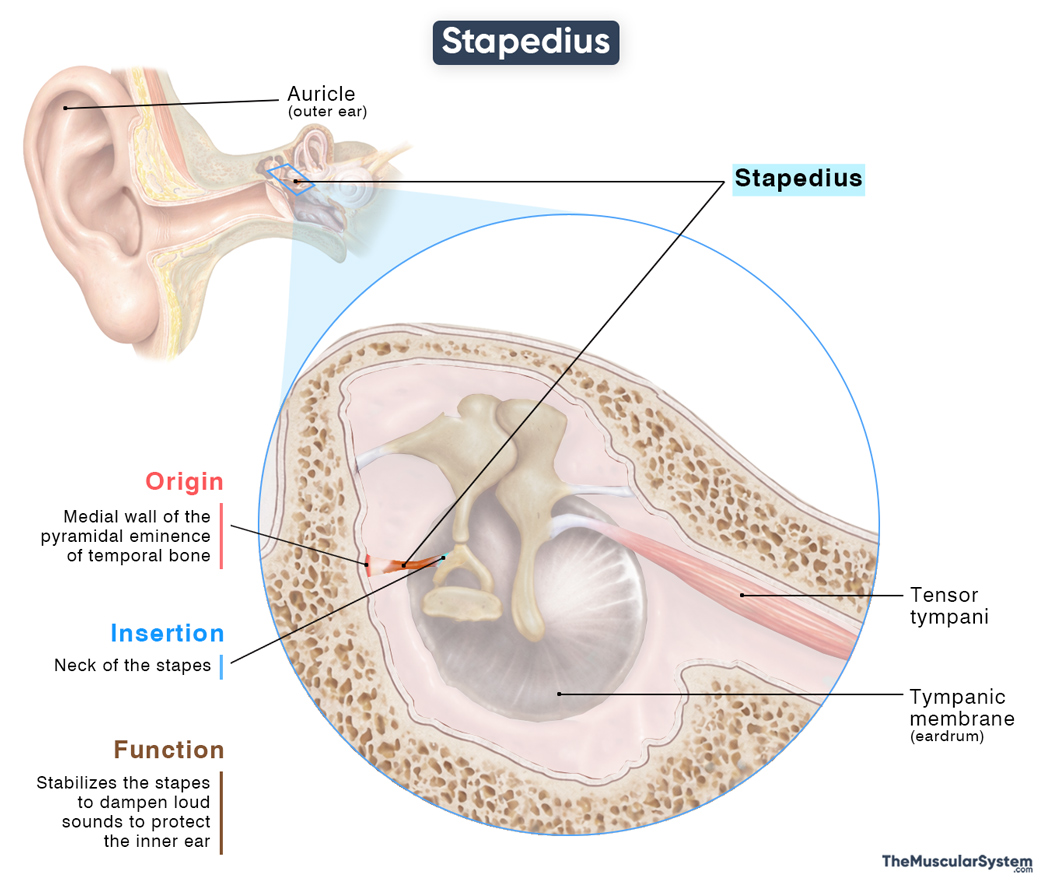
| Origin | The medial wall of the pyramidal eminence of the temporal bone |
| Insertion | Neck of the stapes |
Origin
The stapedius muscle originates from the internal surface of the pyramidal eminence, a hollow projection on the mastoid part of the temporal bone.
Insertion
From its origin, the tendon broadens to form the small muscle belly as the fibers course anteriorly to insert onto the posterior surface of the neck of the stapes bone.
Relations With Surrounding Muscles and Structures
The stapedius lies deep within the posterior portion of the tympanic cavity. It is closely related to several important nerves in the area. Superiorly, it is related to the mastoid segment of the facial nerve as it runs in the facial canal. The chorda tympani passes anterior and slightly superior to it, while the tympanic cavity, the air-filled space in the middle ear, forms its lateral and medial boundaries.
Function
| Action | Stabilizing the stapes and dampening its movement during loud sounds to protect the inner ear |
To understand what the stapedius does, it helps to first understand the functioning of the middle ear ossicles. The stapes is a tiny, stirrup-shaped bone that passes sound vibrations from the middle ear into the inner ear. If the stapes vibrates too strongly, it can send excessive force into the inner ear and potentially cause damage.
The stapedius helps prevent this by stabilizing the stapes and controlling the strength of its movement. It works along with the tensor tympani to produce the stapedius reflex, also called the acoustic reflex. When a loud sound occurs, these muscles contract automatically and reduce how much the ossicles vibrate. This protects the inner ear from sudden bursts of loud noise. The reflex also has a filtering effect: it dampens low-frequency background sounds, which helps you focus on specific sounds more easily in noisy environments.
Because the stapedius plays a vital role in moderating sound transmission, damage or paralysis of the muscle can leave the stapes largely undampened. This may lead to hyperacusis, a hearing disorder where ordinary sounds feel unusually loud and uncomfortable.
Antagonists
Although the stapedius has no true anatomical antagonist, its pull on the stapes produces a mechanical action that is functionally antagonistic to the inward movement generated by the tensor tympani.
Innervation
| Nerve | Nerve to the stapedius (CN VII) |
The nerve to the stapedius muscle, a tiny branch of the facial nerve (CN VII), provides motor innervation to the muscle. So, when there is a loud sound, the brain sends a signal through this nerve to activate the stapedius reflex.
Blood Supply
| Artery | Posterior auricular artery |
Blood supply to the muscle primarily comes from the posterior auricular artery, a branch of the external carotid artery. The anterior tympanic artery, which branches off the mandibular portion of the maxillary artery, also a branch of the external carotid artery, provides additional blood supply.
References
- Stapedius Muscle | Definition, Function & Disorder: Study.com
- Stapedius Muscle: Kenhub.com
- Stapedius Muscle: Radiopaedia.org
- Stapedius: Meddean.LUC.edu
- Stapedius Muscle: IMAIOS.com
- Microsurgical Anatomy of Stapedius Muscle: Anatomy Revisited, Redefined with Potential Impact in Surgeries: PMC.NCBI.NLM.NIH.gov
Della Barnes, an MS Anatomy graduate, blends medical research with accessible writing, simplifying complex anatomy for a better understanding and appreciation of human anatomy.
- Latest Posts by Della Barnes, MS Anatomy
-
Tensor Tympani
- -
Auricularis Posterior
- -
Auricularis Superior
- All Posts


