

Auricularis Superior
By
Della Barnes, an MS Anatomy graduate, blends medical research with accessible writing, simplifying complex anatomy for a better understanding and appreciation of human anatomy.
Last updated:
12/02/2026Della Barnes, MS Anatomy
UX/UI Designer at - AdobeDella Barnes, an MS Anatomy graduate, blends medical research with accessible writing, simplifying complex anatomy for a better understanding and appreciation of human anatomy.
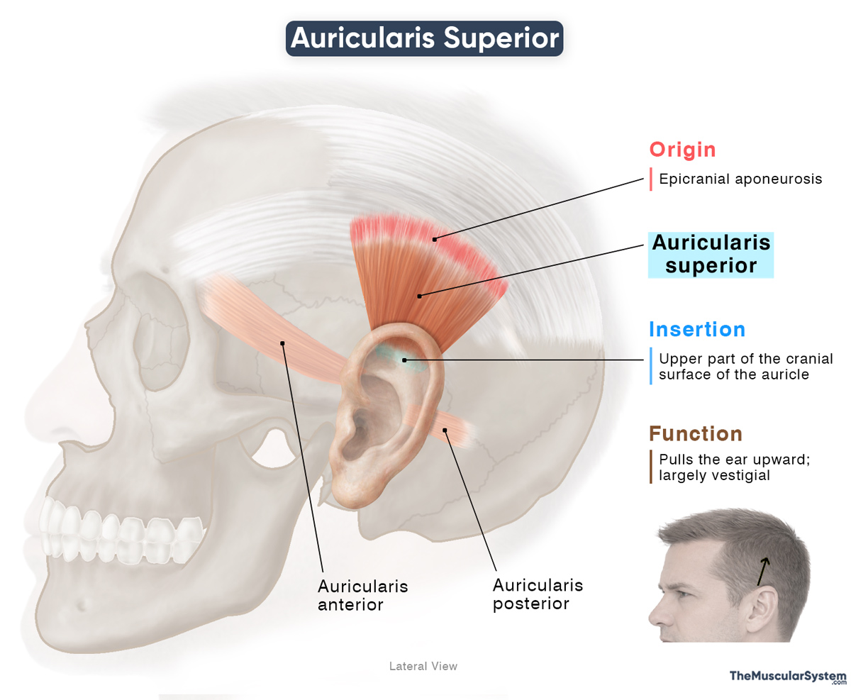
The auricularis superior is a flat, triangular muscle situated above the auricle, or external ear, in the temporal region of the skull. Also known as the superior auricular muscle, it is the largest and most developed of the three auricular muscles, the others being the auricularis anterior and auricularis posterior. Its action is to elevate the auricle, though in most humans this movement is minimal or absent.
Anatomy
Location and Attachments
| Origin | Epicranial aponeurosis |
| Insertion | Upper part of the cranial surface of the auricle |
Origin
The muscle originates as tendinous fibers from the sides of the epicranial aponeurosis in the temporal region of the skull.
Insertion
From their origin, the tendons course downward, converging together as they go, forming a fan-shaped belly. Finally, the muscle inserts via a broad tendon into the upper portion of the cranial (medial) surface of the auricle, in the region just superior to the helix.
Relations With Surrounding Muscles and Structures
It lies between the anterior and posterior auricular muscles and deep to the temporoparietal (superficial temporal) fascia. The muscle is superficial to the temporal fascia and the temporalis muscle, and sits just above the superior part of the auricle.
Near the auricle, its fibers blend and become continuous with the originating fibers of the temporoparietalis muscle.
Function
| Action | Pulling the ear upward |
With its insertion along the upper cranial surface of the auricle, when the superior auricular muscle contracts, it elevates the auricle and pulls it slightly backward.
Although its actions are barely perceptible in humans, the muscle once helped ancestral primates orient their ears toward sound, and in identifying different sounds. In fact, research suggests that the muscle still produces subtle auricular movements during focused listening, but it is generally considered vestigial.
In some rare cases, the muscle may move the auricle more noticeably, allowing certain individuals to move or ‘wiggle’ their ears. However, this does not improve hearing.
Antagonists
The muscle does not have a direct antagonist, because its action is too minor to require an opposing muscle, and the auricle passively returns to its resting position once the muscle relaxes.
Innervation
| Nerve | Temporal branches of the facial nerve |
The muscle receives innervation from the temporal branches of the facial nerve (CN VII), the same nerve that supplies most other facial muscles.
Blood Supply
| Artery | Posterior auricular artery and the anterior auricular branches of the superficial temporal artery |
Its primary blood supply comes from the posterior auricular artery, while the anterior auricular branches of the superficial temporal artery provide additional supply. Both arteries are branches of the external carotid artery.
References
- Auricularis Superior Muscle: Elsevier.com
- Auricularis Superior Muscle: IMAIOS.com
- Superior Auricular Muscle: Radiopaedia.org
- Auricular Muscles: Sciencedirect.com
Della Barnes, an MS Anatomy graduate, blends medical research with accessible writing, simplifying complex anatomy for a better understanding and appreciation of human anatomy.
- Latest Posts by Della Barnes, MS Anatomy
-
Tensor Tympani
- -
Stapedius
- -
Auricularis Posterior
- All Posts


