

Auricularis Anterior
By
Della Barnes, an MS Anatomy graduate, blends medical research with accessible writing, simplifying complex anatomy for a better understanding and appreciation of human anatomy.
Last updated:
12/02/2026Della Barnes, MS Anatomy
UX/UI Designer at - AdobeDella Barnes, an MS Anatomy graduate, blends medical research with accessible writing, simplifying complex anatomy for a better understanding and appreciation of human anatomy.
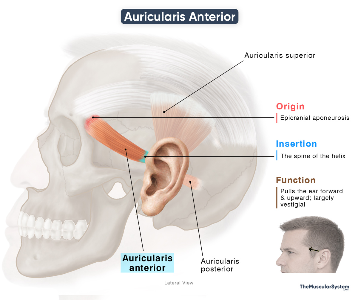
The auricularis anterior is a small, paired muscle located on the sides of the skull, between the eye orbit and the ear. Also known as the anterior auricular muscle, it is one of the three auricular muscles, along with the auricularis superior and auricularis posterior. These muscles primarily assist in producing slight movements of the auricle, although their function in humans is mostly vestigial.
Anatomy
Location and Attachments
| Origin | Epicranial aponeurosis |
| Insertion | The spine of the helix |
Origin
The muscle arises from the lateral portion of the epicranial aponeurosis, the broad tendinous sheet covering the top of the cranium. Additional fibers may originate from the temporal fascia overlying the temporal region.
Insertion
The tendons form a flat, broad belly, with the muscle fibers coursing obliquely downward to insert into the small projection at the anterior base of the helix, known as the spine of the helix. This attachment lies just in front of the concha, close to where the auricle, the external part of the ear, connects to the head.
Relations With Surrounding Muscles and Structures
It is the smallest and most anterior of the three auricularis muscles, lying superficial to the deep temporal fascia. Some of its fibers near the helix intermingle with the temporoparietalis muscle, as it originates from the auricular muscles.
The superficial temporal artery courses deep to the muscle as it travels to supply the auricle.
Function
| Action | Pulling the ear forward |
Because it inserts near the base of the helix, contraction of the anterior auricular muscle pulls the auricle of the ear a little forward and upward. The movement is usually subtle, but in people who can “wiggle” their ears, it becomes more noticeable.
This action has been found to have helped ancestral primates to orient their ears toward sound. Although, with development in other faculties like vision, speech, and greater cognitive processing, humans lost the need to move their ears, and the muscle became vestigial.
Antagonists
There is no functional antagonist muscle, as the movement it produces is minimal, and the ear returns to its resting position passively once the muscle relaxes.
Innervation
| Nerve | Temporal branches of the facial nerve |
Like most of the other facial muscles, it is mainly innervated by the temporal branches of the facial nerve (VII).
Blood Supply
| Artery | Posterior auricular artery, anterior auricular branches of superficial temporal artery |
Blood supply to the muscle comes primarily from the posterior auricular artery, a branch of the external carotid artery. It also receives contributions from the anterior auricular branches of the superficial temporal artery.
References
- Auricularis Anterior Muscle: Elsevier.com
- Auricularis Anterior Muscle: IMAIOS.com
- Auricularis anterior: HealthLine.com
- Anterior Auricular Muscle: Radiopaedia.org
- Auricular Muscles: Sciencedirect.com
Della Barnes, an MS Anatomy graduate, blends medical research with accessible writing, simplifying complex anatomy for a better understanding and appreciation of human anatomy.
- Latest Posts by Della Barnes, MS Anatomy
-
Tensor Tympani
- -
Stapedius
- -
Auricularis Posterior
- All Posts


