

Temporoparietalis
By
Della Barnes, an MS Anatomy graduate, blends medical research with accessible writing, simplifying complex anatomy for a better understanding and appreciation of human anatomy.
Last updated:
09/02/2026Della Barnes, MS Anatomy
UX/UI Designer at - AdobeDella Barnes, an MS Anatomy graduate, blends medical research with accessible writing, simplifying complex anatomy for a better understanding and appreciation of human anatomy.
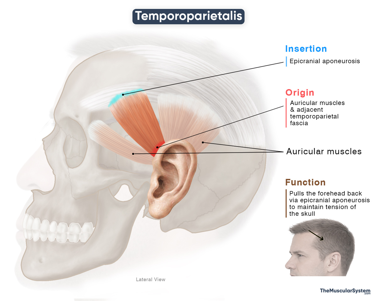
The temporoparietalis is a small, thin, fan-shaped muscle located on the sides of the head, above the ears. Despite often being rather poorly developed, it helps with facial expression and scalp movement.
Anatomy
Location and Attachments
| Origin | The auricular muscles and temporoparietal fascia |
| Insertion | Epicranial aponeurosis |
Origin
The muscle has no bony attachments. It arises from the auricular muscles and the overlying temporoparietal fascia in the region above the outer ear.
Insertion
Its fibers ascend obliquely to insert into the epicranial aponeurosis (galea aponeurotica), which overlies the parietal bone and covers the superior aspect of the cranium.
Relations With Surrounding Muscles and Structures
The muscle lies superficial to the deep temporal fascia, positioned within the temporoparietal fascia layer that covers the temporal fossa. It is continuous with the anterior, superior, and posterior auricular muscles, whose fibers contribute to the origin of the temporoparietalis. It also lies superficial to the anterior portion of the temporalis muscle, which is the primary muscle used for chewing.
Function
| Action | Helps maintain scalp tension |
The muscle does not perform any major action, but assists nearby muscles and fascia in their functions. Through its connection with the epicranial aponeurosis and the occipitofrontalis, its contraction produces a slight posterior and upward tension on the scalp, helping maintain its tautness over the temporal region.
Antagonists
The muscle has no direct antagonists, as it does not generate any primary or isolated movements.
Innervation
| Nerve | Temporal branches of the facial nerve |
Its innervation is the same as that of the nearby facial muscles, supplied by the temporal branches of the facial nerve (CN VII).
Blood Supply
| Artery | Posterior auricular artery |
The posterior auricular artery, a branch of the external carotid artery, provides blood supply to this muscle.
References
- Temporoparietalis Muscle: Elsevier.com
- Temporoparietalis (Anatomy): PrimaryCareNotebook
- Temporoparietalis: Meddean.LUC.edu
Della Barnes, an MS Anatomy graduate, blends medical research with accessible writing, simplifying complex anatomy for a better understanding and appreciation of human anatomy.
- Latest Posts by Della Barnes, MS Anatomy
-
Tensor Tympani
- -
Stapedius
- -
Auricularis Posterior
- All Posts


