

Suboccipital Muscles
By
Della Barnes, an MS Anatomy graduate, blends medical research with accessible writing, simplifying complex anatomy for a better understanding and appreciation of human anatomy.
Last updated:
15/12/2025Della Barnes, MS Anatomy
UX/UI Designer at - AdobeDella Barnes, an MS Anatomy graduate, blends medical research with accessible writing, simplifying complex anatomy for a better understanding and appreciation of human anatomy.
What is the Suboccipital Region
The suboccipital region is the area between the atlas (the first cervical vertebra) below and the external occipital protuberance and inferior nuchal line of the occipital bone above. Within this small compartment lie four paired muscles, referred to as the suboccipital muscles, or suboccipitals, that connect the upper cervical spine to the back of the skull.
These muscles play a vital role in extending, rotating, and stabilizing the atlanto-occipital joint, the articulation between the atlas and the occipital bone.
Names and Anatomy of the Suboccipital Muscles
The suboccipitals are the deepest muscles of the posterior neck, positioned beneath the larger superficial muscles, including the trapezius, splenius, semispinalis, and sternocleidomastoid.
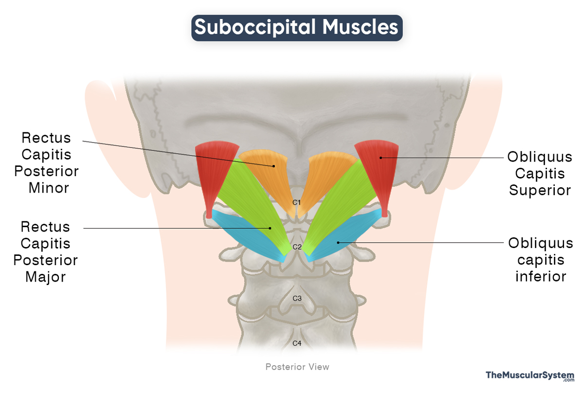
Two of these muscles run obliquely (the obliquus muscles), while the other two run vertically (the rectus muscles). Here is a list of these four muscles with their attachments and primary functions:
| Name | Origin | Insertion | Action |
|---|---|---|---|
| Obliquus Capitis Superior The smallest and most lateral muscle in the group | Transverse process of the atlas (C1 vertebra) | Posterior surface of the occipital bone, between the inferior and superior nuchal lines | Extending and laterally flexing the neck, and stabilizing the atlanto-occipital joint |
| Obliquus Capitis Inferior* The largest muscle in the group | Spinous process of the axis (C2 vertebra) | Transverse process of the atlas (C1 vertebra) | Extending, ipsilaterally rotating, and stabilizing the head and neck at the atlanto-axial joint |
| Rectus Capitis Posterior Major | Spinous process of the axis (C2 vertebra) | The lateral half of the inferior nuchal line of the occipital bone | Extending and laterally rotating the neck at the atlanto-occipital joint |
| Rectus Capitis Posterior Minor The most medial muscle in the group | Posterior tubercle of the atlas (C1 vertebra) | The medial half of the inferior nuchal line of the occipital bone | Extending the neck at the atlanto-occipital joint (weakly) |
Innervation: All four suboccipital muscles are innervated by the suboccipital nerve, which is the primary dorsal ramus of the first cervical (C1) spinal nerve.
Blood Supply: Like their innervation, the four muscles receive blood supply from the same source, the vertebral artery and the deep branch of the descending occipital artery. The vertebral artery branches off the subclavian artery, while the occipital artery is a branch of the external carotid artery.
Relations with Surrounding Structures
Suboccipital Triangle
The suboccipital triangle is an important anatomical space at the back of the neck, bordered by three of the four suboccipital muscles.
- Upper medial boundary: Rectus capitis posterior major
- Upper lateral boundary: Obliquus capitis superior
- Lower lateral boundary: Obliquus capitis inferior
Its floor is formed by the posterior atlantooccipital membrane and the posterior arch of the C1 vertebra (atlas).
This triangle provides passage for vital neurovascular structures, including the vertebral artery, suboccipital nerve, and suboccipital venous plexus, all of which are essential for neurovascular supply to the back of the head and neck.
Spinal Dura Mater
The rectus capitis posterior minor forms an indirect connective-tissue attachment to the spinal dura mater via the myodural bridge. Anatomical studies show that the rectus capitis posterior major and the obliquus capitis inferior may also form similar links. Due to this connection, stress and tension in these muscles are often associated with headaches that start at the back of the neck.
References
- The Suboccipital Muscles: TeachMeAnatomy.info
- Anatomy, Head and Neck, Suboccipital Muscles: NCBI.NLM.NIH.gov
- Suboccipital Muscles: Kenhub.com
- Suboccipital Muscle Group: Radiopaedia.org
- Suboccipital Muscles: Elsevier.com
- Anatomy of the Suboccipital Region: Osmosis.org
Della Barnes, an MS Anatomy graduate, blends medical research with accessible writing, simplifying complex anatomy for a better understanding and appreciation of human anatomy.
- Latest Posts by Della Barnes, MS Anatomy
-
Tensor Tympani
- -
Stapedius
- -
Auricularis Posterior
- All Posts


