

Rectus Capitis Posterior Major
By
Della Barnes, an MS Anatomy graduate, blends medical research with accessible writing, simplifying complex anatomy for a better understanding and appreciation of human anatomy.
Last updated:
24/11/2025Della Barnes, MS Anatomy
UX/UI Designer at - AdobeDella Barnes, an MS Anatomy graduate, blends medical research with accessible writing, simplifying complex anatomy for a better understanding and appreciation of human anatomy.
What is the Rectus Capitis Posterior Major
The rectus capitis posterior major is a small, paired muscle located at the upper back of the neck. It assists in movements such as extension and rotation of the head and neck. This muscle is part of the suboccipital group, which also includes the rectus capitis posterior minor, obliquus capitis superior, and obliquus capitis inferior.
Anatomy
Location and Attachments
| Origin | Spinous process of the axis bone |
| Insertion | The lateral half of the inferior nuchal line of the occipital bone |
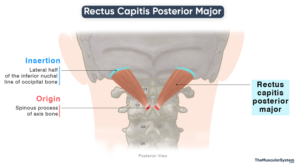
Origin
The muscle originates from the outer surface of the spinous process of the axis (C2 vertebra) via a thin tendon.
Insertion
The originating fibers broaden to form a triangular muscle belly that ascends laterally, crossing the atlas (first cervical vertebra, C1). It has a broad insertion on the lateral half of the inferior nuchal line on the posterior surface of the occipital bone. Additionally, a few fibers attach just below the inferior nuchal line on the occipital bone.
Relations With Surrounding Muscles and Structures
The muscle lies medial and deep to the obliquus capitis superior, while it is lateral and superficial to the rectus capitis posterior minor. Together, these three muscles form the suboccipital triangle, with the rectus capitis posterior major forming its upper medial boundary. The suboccipital triangle is an anatomical space that allows passage to the vertebral and suboccipital neurovascular structures.
Studies have shown a soft tissue connection (myodural bridge) between the muscle and the cervical dura mater in the atlantoaxial region. It is believed to help stabilize the spinal dura mater, although further research is necessary to confirm this relationship.
Function
| Action | Extending and ipsilaterally rotating the neck at the atlanto-occipital joint |
- When the rectus capitis posterior major contracts bilaterally, it extends the atlanto-occipital joint, producing neck extension—such as when you lift your head to look upward.
- In contrast, unilateral contraction rotates the head ipsilaterally, meaning toward the same side as the contracting muscle.
The muscle works in coordination with the obliquus capitis inferior and the splenius capitis, both of which assist in neck rotation.
Together with the other suboccipital muscles, it also serves an important postural function, stabilizing the atlanto-occipital joint and helping maintain the head’s position on the neck.
Antagonists
The longus capitis and rectus capitis anterior, which flex the head at the atlanto-occipital joint, oppose this muscle in neck extension. Additionally, the sternocleidomastoid acts as a functional antagonist during rotation, since it turns the head contralaterally, opposite to the ipsilateral rotation produced by the rectus capitis posterior major.
Innervation
| Nerve | Suboccipital nerve |
Like the other suboccipital muscles, it is innervated by the suboccipital nerve, arising from the posterior ramus of the first cervical spinal nerve (C1).
Blood Supply
| Artery | Vertebral artery and deep descending occipital artery |
Its blood supply comes from the vertebral artery, a branch of the subclavian artery, and the deep branch of the descending occipital artery, which originates from the external carotid artery.
References
- Rectus Capitis Posterior Major Muscle: Kenhub.com
- Rectus Posterior Major Capitis Muscle: Elsevier.com
- Rectus Capitis Posterior Major: TeachMeAnatomy.info
- Rectus Capitis Posterior Major: GetBodySmart.com
Della Barnes, an MS Anatomy graduate, blends medical research with accessible writing, simplifying complex anatomy for a better understanding and appreciation of human anatomy.
- Latest Posts by Della Barnes, MS Anatomy
-
Tensor Tympani
- -
Stapedius
- -
Auricularis Posterior
- All Posts


