

Laryngeal Muscles
By
Della Barnes, an MS Anatomy graduate, blends medical research with accessible writing, simplifying complex anatomy for a better understanding and appreciation of human anatomy.
Last updated:
28/01/2026Della Barnes, MS Anatomy
UX/UI Designer at - AdobeDella Barnes, an MS Anatomy graduate, blends medical research with accessible writing, simplifying complex anatomy for a better understanding and appreciation of human anatomy.
The larynx, or voice box, is a flexible, hollow organ located in the upper front part of the neck. It is primarily made up of several paired and unpaired cartilages that serve as points of attachment for the six intrinsic laryngeal muscles. These muscles adjust the position, tension, and shape of the vocal folds (vocal cords) to help with phonation, pitch control, and also the protection of the airways. Additionally, the larynx has several extrinsic muscles that help with its actions.
Names and Anatomy of the Muscles of the Larynx
When discussing laryngeal muscles, primary consideration is given to the intrinsic muscles that originate within the larynx and act on its structures.
Intrinsic Laryngeal Muscles
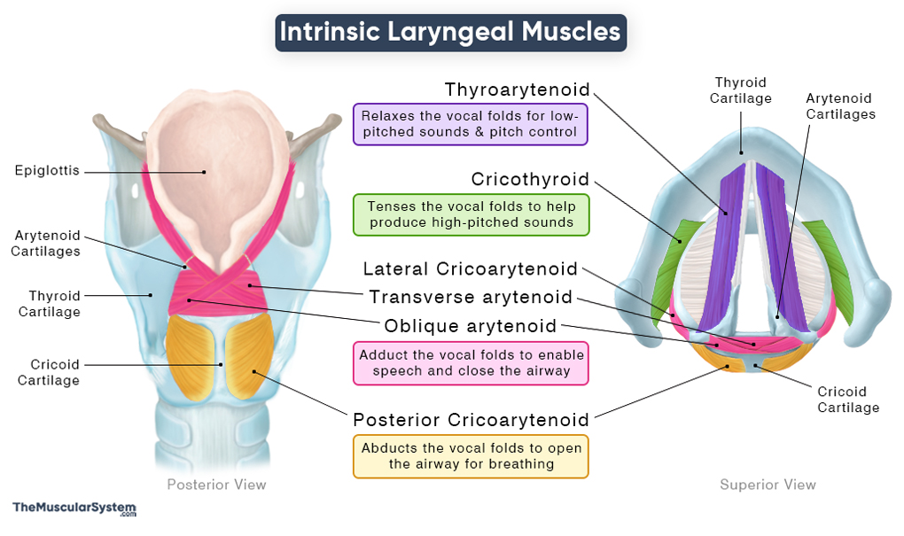
There are five paired and one unpaired intrinsic muscle that make up the group of intrinsic muscles of the larynx. These muscles can be grouped based on their action on the vocal folds. Here is a list of their names with their basic anatomy and actions:
| Name | Origin | Insertion | Action | Innervation | Blood Supply |
|---|---|---|---|---|---|
| Tensors/Relaxors of the Vocal Folds Includes a pair of antagonistic muscles that adjust vocal fold tension for pitch modulation. As they do not directly produce phonation, their dysfunction impairs pitch without causing loss of voice. | |||||
| Cricothyroid | Anterolateral aspect of the cricoid cartilage | Straight Part: Inferior border of the lamina of the thyroid cartilage Oblique Part: Inferior cornu (horn) of the thyroid cartilage | Tensing the vocal folds to help with high-pitched vocalization | External laryngeal branch of the superior laryngeal nerve | Cricothyroid artery |
| Thyroarytenoid | infero-anterior surface of the thyroid cartilage, between the two lamina | anterior and lateralsurfaces of the arytenoid cartilage | Relaxing the vocal cords to help produce low-pitch sounds | Recurrent laryngeal nerve | Superior and inferior thyroid arteries |
| Abductors of the Vocal Folds Includes the only muscle that abducts the vocal folds to open the rima glottidis, allowing airflow during breathing; dysfunction may be life-threatening due to compromised airway patency. | |||||
| Posterior Cricoarytenoid | Posterior surface of the cricoid cartilage | Posterior and superior surfaces of the muscular process of arytenoid cartilage | Abducting the vocal folds to help with breathing and vocalization | Recurrent laryngeal nerve | Superior and inferior thyroid arteries |
| Adductors of the Vocal Folds Includes the primary muscles for speech, which adduct the vocal folds to close the rima glottidis for phonation and airway protection; dysfunction may lead to loss of voice along with an increased risk of aspiration. | |||||
| Lateral Cricoarytenoid | Superior edge of the cricoid arch on the cricoid cartilage | Anterior surface of the muscular process of the ipsilateral arytenoid cartilage | — Adducting the vocal folds to help with vocalization — Helping protect the airways by keeping it closed during swallowing | Recurrent laryngeal nerve | Superior and inferior thyroid arteries |
| Oblique arytenoid | The muscular process of the arytenoid cartilage | The apex and posterior surface of the arytenoid cartilage on the contralateral side | — Working as a sphincter to close the laryngeal opening during swallowing and coughing — Assisting in vocalization | Recurrent laryngeal nerve | Superior and inferior thyroid arteries |
| Transverse arytenoid | Posterior surface of the muscular process and the lateral margin of the arytenoid cartilage on one side | The corresponding area on the contralateral arytenoid cartilage | — Helping close the laryngeal opening to prevent aspiration of food and water — Assisting in vocalization | Recurrent laryngeal nerve | Superior and inferior thyroid arteries |
An interesting point to notice about the cricothyroid muscle is that both its nerve and blood supply come from different sources compared to the rest of the intrinsic muscles. It is because the muscle is derived from the 4th pharyngeal arch during embryonic development, whereas the other five muscles arise from the 6th pharyngeal arch.
A mnemonic to easily remember the nerve supply to these muscles is “SCAR”:
- S: Superior laryngeal nerve (via its external laryngeal branch)
- C: Cricothyroid muscle
- A: All other intrinsic muscles
- R: Recurrent laryngeal nerve
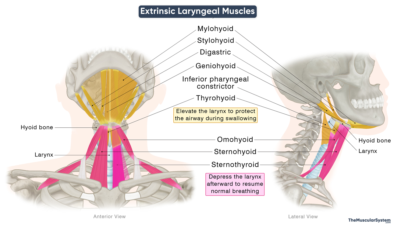
Extrinsic Laryngeal Muscles
The extrinsic laryngeal muscles are muscles that originate outside the larynx and insert into the laryngeal cartilages or hyoid bone. Rather than directly acting on the vocal folds, their primary role is to move the larynx as a whole, which is essential for swallowing, phonation, and pitch control.
They come from different groups of neck and facial muscles, and are divided into two functional groups:
| Muscles that elevate the larynx | |
| Pharyngeal muscles | Inferior pharyngeal constrictors |
| Infrahyoid muscles | Thyrohyoid |
| Suprahyoid muscles | Mylohyoid, stylohyoid, digastric, geniohyoid |
| Muscles that depress the larynx | |
| Infrahyoid muscles | Sternothyroid, omohyoid, sternohyoid |
The muscles that elevate the larynx narrow the laryngeal opening during swallowing to protect the airway and guide food and liquids into the pharynx. The opposing muscles depress the larynx afterward to resume normal breathing.
References
- Laryngeal Muscles: TeachMeAnatomy.info
- Anatomy, Head and Neck: Laryngeal Muscles: NCBI.NLM.NIH.gov
- Muscles of the Larynx: Kenhub.com
- Laryngeal Anatomy: Muscles and Innervation: OpenAnesthesia.org
- Laryngeal Muscles: IMAIOS.com
Della Barnes, an MS Anatomy graduate, blends medical research with accessible writing, simplifying complex anatomy for a better understanding and appreciation of human anatomy.
- Latest Posts by Della Barnes, MS Anatomy
-
Tensor Tympani
- -
Stapedius
- -
Auricularis Posterior
- All Posts


