

Sternothyroid
By
Della Barnes, an MS Anatomy graduate, blends medical research with accessible writing, simplifying complex anatomy for a better understanding and appreciation of human anatomy.
Last updated:
08/12/2025Della Barnes, MS Anatomy
UX/UI Designer at - AdobeDella Barnes, an MS Anatomy graduate, blends medical research with accessible writing, simplifying complex anatomy for a better understanding and appreciation of human anatomy.
What is the Sternothyroid
Sternothyroid is a broad, paired strap muscle at the lower front of the neck. It forms the infrahyoid muscle group together with the thyrohyoid, sternohyoid, and omohyoid muscles. Its functions include stabilizing the hyoid bone and helping with swallowing.
Anatomy
Location and Attachments
| Origin | Back of the manubrium and first costal cartilage |
| Insertion | The oblique line of the thyroid cartilage |
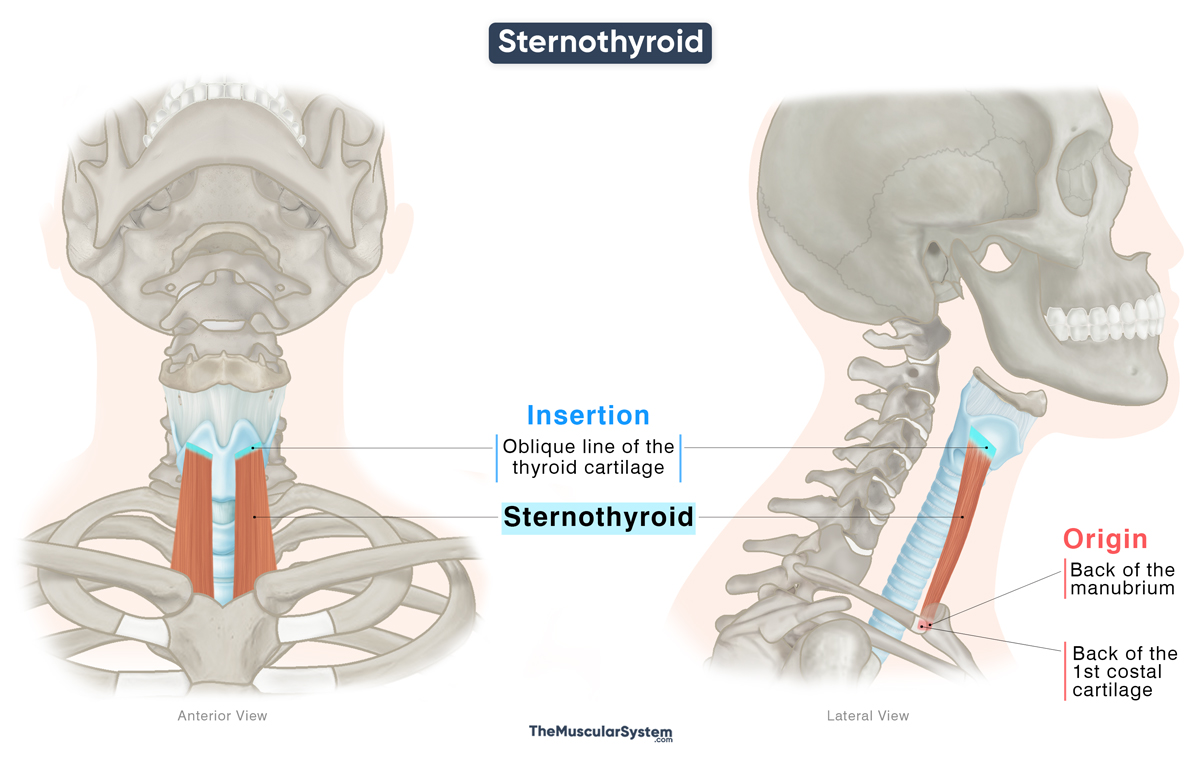
Origin
The muscle originates from the back of the manubrium, the top part of the sternum. The point of origin extends to include the posterior surface of the first costal cartilage.
Insertion
At their origins on the posterior surface of the manubrium, the left and right sternothyroid muscles lie close to each other on either side of the midline. As they ascend vertically and slightly laterally, the muscle fibers diverge, forming distinct muscle bellies. They each insert into the lamina of the thyroid cartilage, along the oblique line.
Relations With Surrounding Muscles and Structures
With the other infrahyoid muscles
The point of origin of this muscle lies just below that of the sternohyoid. As the muscle bellies course upward, the sternothyroid lies partly medial and deep to the sternohyoid, appearing shorter and broader in comparison. The superior belly of the omohyoid muscle also passes superficial to the sternothyroid.
The point of insertion of the sternothyroid lies along the oblique line of the thyroid cartilage, at a position directly below the region from which the thyrohyoid muscle arises.
With neurovascular and other structures
The left and right lobes of the thyroid gland lie in front of the corresponding sternothyroid muscles. The inferior pharyngeal constrictor muscle and the cervical portion of the trachea are located posteriorly.
The muscle has several neurovascular relations. On the right, it lies anterior to the brachiocephalic trunk, while on the left, it is anterior to the common carotid artery and the brachiocephalic vein. It also lies superficial to the external laryngeal nerve and superior thyroid artery.
Variations
Accessory slips of muscle fibers may extend from the sternothyroid to attach to the thyrohyoid. It may also vary in size and, in rare cases, may be absent on one or both sides.
Function
| Action | Depressing the hyoid bone and larynx to help with breathing and vocalization |
During swallowing, the suprahyoid muscles lift the hyoid to help close the laryngeal opening, preventing food from entering the airways. After swallowing, the sternothyroid, together with the other infrahyoid muscles, helps depress the hyoid bone and larynx, returning them to their resting position so breathing can resume.
When the hyoid bone is held in place by the suprahyoid muscles, this muscle helps elevate the larynx, which helps in producing low-pitched sounds.
Antagonists
The thyrohyoid muscle has no single direct antagonist, but as an infrahyoid muscle that depresses the hyoid, its action is functionally opposed by the suprahyoid muscles, the mylohyoid, stylohyoid, digastric, and geniohyoid, which elevate the hyoid.
Innervation
| Nerve | Ansa cervicalis (C1-C3) |
The muscle is innervated by the ansa cervicalis, a nerve loop derived from the anterior rami of the first to third cervical (C1-C3) spinal nerves.
Blood Supply
| Artery | Infrahyoid branch of the superior thyroid artery |
Primary blood supply to the muscle comes from the infrahyoid branch of the superior thyroid artery, with additional supply from the lingual artery. Both these arteries arise from the external carotid artery.
References
- Sternothyroid Muscle: Kenhub.com
- Sternothyroid: TeachMeAnatomy.info
- Sternothyroid: HealthLine.com
- Sternothyroid Muscle: Elsevier.com
Della Barnes, an MS Anatomy graduate, blends medical research with accessible writing, simplifying complex anatomy for a better understanding and appreciation of human anatomy.
- Latest Posts by Della Barnes, MS Anatomy
-
Tensor Tympani
- -
Stapedius
- -
Auricularis Posterior
- All Posts


