

Inferior Pharyngeal Constrictor
By
Della Barnes, an MS Anatomy graduate, blends medical research with accessible writing, simplifying complex anatomy for a better understanding and appreciation of human anatomy.
Last updated:
19/12/2025Della Barnes, MS Anatomy
UX/UI Designer at - AdobeDella Barnes, an MS Anatomy graduate, blends medical research with accessible writing, simplifying complex anatomy for a better understanding and appreciation of human anatomy.
The inferior pharyngeal constrictor is the lowermost of the three pharyngeal constrictors, located in the neck, on the sides and back of the pharynx. It is the thickest and most superficial of the three constrictor muscles. As a constrictor muscle, it helps propel food down to the esophagus during swallowing.
Anatomy
Location and Attachments
| Origin | The oblique line of the thyroid cartilage and the cricoid cartilage |
| Insertion | Median pharyngeal raphe |
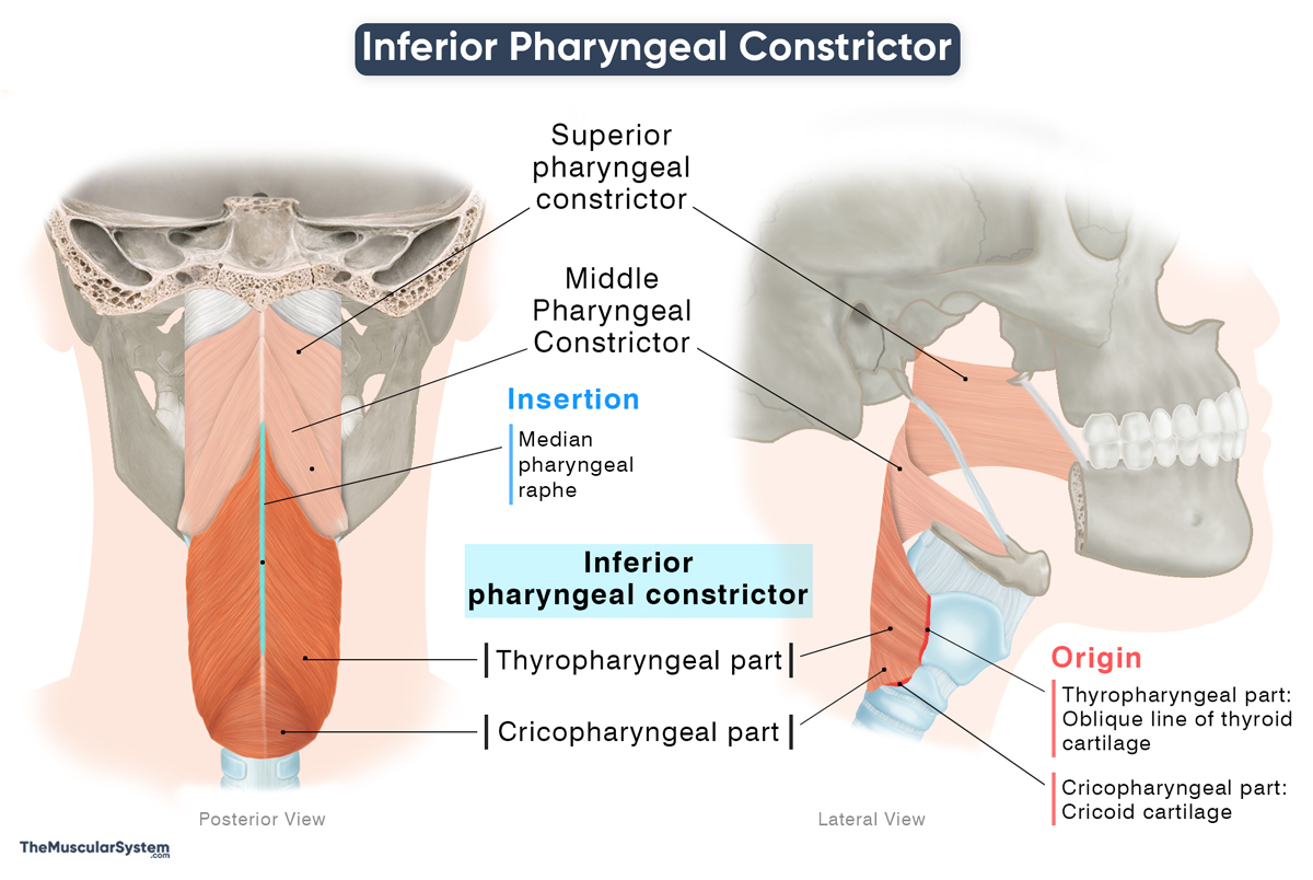
The muscle originates from two primary points and is divided into two parts accordingly.
Origin
Thyropharyngeal part: The superior fibers that comprise most of the muscle originate from the oblique line of the thyroid cartilage. Some fibers may also arise from the posterior borders and the inferior horns of the cartilage.
Cricopharyngeal part: The smaller and inferior portion of the muscle originates from the cricoid cartilage. The point of origin lies in the gap between the small laryngeal muscle, cricothyroid, and the point where the cricoid cartilage articulates with the inferior horn of the thyroid cartilage. The fibers of this part are further divided into the pars oblique (superior) and pars fundiformis (inferior) based on their orientation.
Insertion
The fibers of the thyropharyngeal part and the pars oblique portion of the cricopharyngeal part course obliquely backward and medially to meet their contralateral fibers. They insert into the median pharyngeal raphe like the rest of the pharyngeal constrictors.
The lower fibers of the pars fundiformis portion course horizontally, blending and becoming continuous with the upper esophageal sphincter.
Relations With Surrounding Muscles and Structures
Anterior and Posterior Relations
The inferior constrictor lies anterior to the cervical vertebral compartment, from which it is separated by the buccopharyngeal fascia. This fascial layer covers the posterior surface of the muscle and forms a boundary between the pharynx and the retropharyngeal space.
The muscle contributes to the sides and back of the muscular wall of the lower pharynx and lies superior to the esophagus.
Superior Relations
Superiorly, the inferior constrictor is related to the middle pharyngeal constrictor. The upper fibers of the inferior constrictor overlap the lower fibers of the middle constrictor, continuing the overlapping arrangement between the middle and superior constrictors above.
Inferior Relations
As it blends with the muscular wall of the esophagus, the cricopharyngeal part forms the uppermost sphincteric region of the esophagus, marking the transition between the pharynx and the esophageal lumen.
Lateral Relations
Laterally, the inferior pharyngeal constrictor is related to the superior poles of the thyroid gland, the common carotid artery, and the sternothyroid muscle. The external branch of the superior laryngeal nerve runs along the outer surface of the thyropharyngeal part before piercing the muscle to reach the larynx, making it an important neurovascular relation in this region.
Since the inferior pharyngeal constrictor consists of two parts with differing fiber orientations, it creates areas of relative weakness in the posterior pharyngeal wall:
- Killian’s triangle: A triangular gap between the thyropharyngeal and cricopharyngeal parts, it is a potential site for herniation of the pharyngeal wall, leading to a Zenker’s diverticulum.
- Laimer’s triangle: A second, less prominent weak area located between the lower edge of the cricopharyngeal part and the upper esophagus. It is the potential location for a Laimer’s diverticula.
Function
| Action | Constricting the lowest portion of the pharynx to propel the bolus into the esophagus |
The muscle plays a vital role during the pharyngeal phase of swallowing. Once the middle pharyngeal constrictor pushes the chewed food (bolus) down, the thyropharyngeal part of the muscle contracts, and the cricopharyngeus relaxes to allow it to move into the esophagus.
After swallowing, the thyropharyngeus relaxes, and the cricopharyngeus resumes tonic contraction. The cricopharyngeus forms the main musculature of the upper esophageal sphincter (UES). Its steady contraction keeps the junction between the pharynx and esophagus closed, so food doesn’t reflux upward and air doesn’t enter the esophagus.
Functionally, the two parts are built for their roles: the thyropharyngeal fibres are mostly fast-twitch (Type II) to produce quick, strong contractions during swallowing, while the cricopharyngeal fibres are mainly slow-twitch (Type I) to maintain the constant tone needed for the UES.
Antagonists
The inferior constrictor does not have any antagonists because the pharyngeal constrictors work mainly by sequential coordination, not by opposition. These muscles work together by alternatively turning on and off rather than in opposing pairs.
Innervation
| Nerve | pharyngeal plexus (vagus nerve) |
Both parts of the muscle are innervated by the pharyngeal plexus of the vagus nerve (CN X). Two additional branches of the vagus nerve, the recurrent and superior laryngeal nerves, may provide additional innervation to the cricopharyngeus part.
Blood Supply
| Artery | Ascending pharyngeal and inferior thyroid arteries |
Its blood supply comes from the ascending pharyngeal artery, a branch of the external carotid artery. The muscular branches of the inferior thyroid artery provide additional supply.
References
- Inferior Pharyngeal Constrictor: TeachMeAnatomy.info
- Inferior Pharyngeal Constrictor Muscle: Radiopaedia.org
- Inferior Pharyngeal Constrictor: Kenhub.com
- Anatomy, Head and Neck: Pharyngeal Muscles: NCBI.NLM.NIH.gov
- Thyropharyngeal Part of Inferior Pharyngeal Constrictor: Elsevier.com
- Cricopharyngeal Part of Inferior Pharyngeal Constrictor: Elsevier.com
Della Barnes, an MS Anatomy graduate, blends medical research with accessible writing, simplifying complex anatomy for a better understanding and appreciation of human anatomy.
- Latest Posts by Della Barnes, MS Anatomy
-
Tensor Tympani
- -
Stapedius
- -
Auricularis Posterior
- All Posts


