

Middle Pharyngeal Constrictor
By
Della Barnes, an MS Anatomy graduate, blends medical research with accessible writing, simplifying complex anatomy for a better understanding and appreciation of human anatomy.
Last updated:
19/12/2025Della Barnes, MS Anatomy
UX/UI Designer at - AdobeDella Barnes, an MS Anatomy graduate, blends medical research with accessible writing, simplifying complex anatomy for a better understanding and appreciation of human anatomy.
What is the Middle Pharyngeal Constrictor
The middle pharyngeal constrictor is a paired, fan-shaped, sheet muscle in the neck, contributing to the wall of the pharynx. Being one of the three constrictors of the pharynx, this muscle coordinates with the superior and inferior constrictors to assist in swallowing.
Anatomy
Location and Attachments
| Origin | The greater and lesser horns of the hyoid bone, and the stylohyoid ligament |
| Insertion | Median pharyngeal raphe |
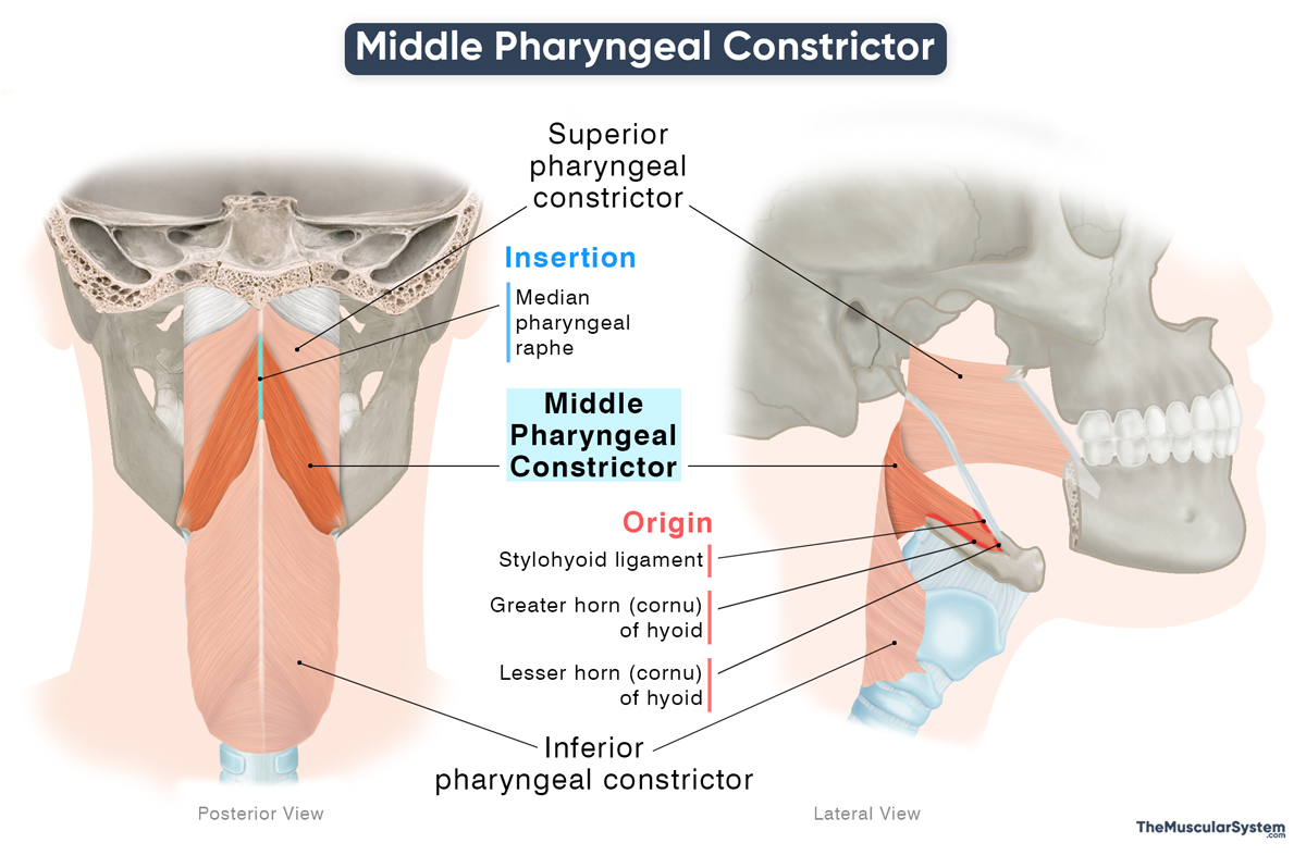
Origin
Like the superior constrictor, the middle constrictor also has multiple points of origin, which divide the muscle into two parts.
- Ceratopharyngeal part: The entire length of the upper surface of the greater horn, or cornu, of the hyoid bone.
- Chondropharyngeal part: The back and sides of the lesser horn of the hyoid, and the lower border of the stylohyoid ligament.
Insertion
From their points of origin, the fibers course posteriorly as they fan out and approach their insertion along the middle portion of the pharyngeal raphe.
The fibers arising from the lesser horn of the hyoid bone and the stylohyoid ligament pass posteriorly and upward, reaching the lower border of the superior pharyngeal constrictor. Some of the fibers originating from the greater horn run horizontally backward, while the remaining fibers descend as they course posteriorly toward the superior fibers of the inferior pharyngeal constrictor.
Relations With Surrounding Muscles and Structures
Anterior and Posterior Relations
The muscle lies posterior to the hyoglossus and other oral floor muscles. At its origin, it sits medial to the hyoglossus before curving back toward the pharyngeal raphe. It forms the middle part of the lateral and posterior pharyngeal wall.
Posteriorly, it is separated from the prevertebral muscles, including the longus capitis, longus colli, and rectus capitis, by the buccopharyngeal fascia and the underlying retropharyngeal space, a potential space behind the pharynx.
As its fibers fan out in the back, they blend with fibers of both the superior and inferior constrictors.
Superior Relations
The upper border of the middle constrictor lies superficial to the lower border of the superior constrictor. The interval between these two muscles forms a small but clinically important passage. The stylopharyngeus muscle, as well as the glossopharyngeal (CN IX) and lingual nerves, pass through this opening. The stylohyoid ligament also passes through the gap as it approaches the pharynx.
Inferior Relations
The lower border of the muscle is deep to the upper border of the inferior constrictor. The different fiber orientations of these two muscles create another natural opening that allows passage to the internal laryngeal nerve, the superior laryngeal artery, and vein.
Lateral Relations
Laterally, the lingual artery courses across the outer surface of the muscle on its way to the tongue. By forming the posterior wall of the pharynx, the muscle also contributes to the medial boundary of the parapharyngeal space, a pyramid-shaped potential space on either side of the pharynx.
Function
| Action | Constricting the middle portion of the pharynx to help propel the bolus down during swallowing |
Along with the other two constrictors, this muscle plays a vital role in the pharyngeal phase of swallowing, contributing to the peristaltic sequence that propels the chewed food (bolus) through the digestive tract.
As the superior constrictor moves the bolus down to the middle portion of the pharynx, this muscle contracts to propel it further downward toward the esophagus.
Antagonists
The middle constrictor has no true antagonist. During swallowing, the muscles of the pharynx work in a synchronized wave-like pattern that moves the bolus downward, so muscles do not function in opposing pairs.
Innervation
| Nerve | Pharyngeal plexus (vagus nerve) |
The muscle receives motor innervation from the pharyngeal plexus via the pharyngeal branch of the vagus nerve (CN X).
Blood Supply
| Artery | The pharyngeal trunk of the ascending pharyngeal artery and the tonsillar artery |
It receives its blood supply from the same sources as the superior constrictor, namely the pharyngeal trunk of the ascending pharyngeal artery and the tonsillar branch of the facial artery, both arising from the external carotid artery.
References
- Middle Pharyngeal Constrictor: Kenhub.com
- Middle Pharyngeal Constrictor: TeachMeAnatomy.info
- Middle Pharyngeal Constrictor Muscle: Radiopaedia.org
- Middle Pharyngeal Constrictor: Elsevier.com
- Anatomy, Head and Neck: Pharyngeal Muscles: NCBI.NLM.NIH.gov
- Middle Pharyngeal Constrictor: IMAIOS.com
Della Barnes, an MS Anatomy graduate, blends medical research with accessible writing, simplifying complex anatomy for a better understanding and appreciation of human anatomy.
- Latest Posts by Della Barnes, MS Anatomy
-
Tensor Tympani
- -
Stapedius
- -
Auricularis Posterior
- All Posts


