

Stylohyoid
By
Della Barnes, an MS Anatomy graduate, blends medical research with accessible writing, simplifying complex anatomy for a better understanding and appreciation of human anatomy.
Last updated:
29/11/2025Della Barnes, MS Anatomy
UX/UI Designer at - AdobeDella Barnes, an MS Anatomy graduate, blends medical research with accessible writing, simplifying complex anatomy for a better understanding and appreciation of human anatomy.
What is the Stylohyoid
The stylohyoid is a narrow, paired muscle in front of the neck. It belongs to the group of suprahyoid muscles along with the mylohyoid, digastric, and geniohyoid muscles. The muscle plays an important role in tongue control during chewing and swallowing.
Anatomy
Location and Attachments
| Origin | Back of the temporal styloid process |
| Insertion | Body of the hyoid bone |
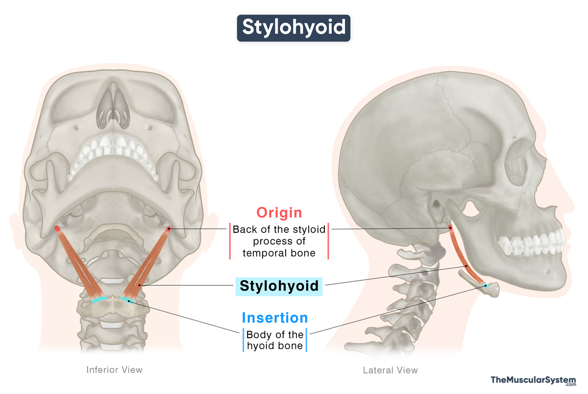
Origin
The muscle originates via a narrow tendon from the temporal styloid process, a thin, pointed projection of the temporal bone that extends downward and slightly forward. The point of origin lies near the base of the process, at its posterior surface.
Insertion
The muscle fibers descend anteriorly and laterally to insert into the superior surface of the body of the hyoid bone, at the junction where it meets the greater cornu.
Relations With Surrounding Muscles and Structures
The stylohyoid runs slightly in front and above the posterior belly of the digastric as it descends toward its insertion on the hyoid bone. Near its distal end, it divides into two slips that pass around the intermediate tendon of the digastric, then reunite to form a single belly before attaching to the hyoid.
The muscle has several important neurovascular relations along its course:
- Deep to it lie the internal carotid artery and the accessory and hypoglossal nerves.
- Inferiorly, it is related to the second part of the lingual artery and the facial artery.
- Laterally superficial to the muscle lie the external carotid artery, the facial vein, and the facial nerve.
- The submandibular gland lies inferiorly behind it.
Variations
Studies show the muscle varies considerably among individuals; it may insert partly into the mylohyoid or omohyoid muscles, its belly may be split into two portions, or the muscle may be absent on one or both sides.
Function
| Action | Elevating the hyoid bone to help with swallowing |
The primary function of the muscle is to pull the hyoid bone posteriorly upward, which in turn lifts the base of the tongue and lengthens the floor of the mouth. These movements are essential during swallowing, especially during the early stages, as they help push the chewed food (bolus) toward the back of the oral cavity and the throat.
As it adds gentle tension to the floor of the oral cavity, the muscle also helps keep the upper airway open during respiration.
When the infrahyoid muscles hold the hyoid bone steady, the stylohyoid works with the other suprahyoid muscles to help depress the mandible, which lowers the jaw to open the mouth wide. Together with these muscles, it also contributes to small movements involved in neck flexion and in producing higher-pitched sounds.
Antagonists
As part of the suprahyoid group, the infrahyoid muscles, the sternohyoid, omohyoid, sternothyroid, and thyrohyoid, can be considered its antagonists because they depress the hyoid bone.
Innervation
| Nerve | Stylohyoid branch of the facial nerve (CN VII) |
The muscle is innervated by the stylohyoid branch of the facial nerve, also known as the seventh cranial nerve (CN VII).
Blood Supply
| Artery | Facial, occipital, and posterior auricular arteries |
Blood supply to this muscle comes from branches of the facial, occipital, and posterior auricular arteries, all arising from the external carotid artery.
References
- Stylohyoid Muscle: Kenhub.com
- Anatomy, Head and Neck, Stylohyoid Muscle: NCBI.NLM.NIH.gov
- The Stylohyoid Muscle: TeachMeAnatomy.info
- Stylohyoid Muscle: IMAIOS.com
- Stylohyoid Muscle: Elsevier.com
Della Barnes, an MS Anatomy graduate, blends medical research with accessible writing, simplifying complex anatomy for a better understanding and appreciation of human anatomy.
- Latest Posts by Della Barnes, MS Anatomy
-
Tensor Tympani
- -
Stapedius
- -
Auricularis Posterior
- All Posts


