

Auricularis Posterior
By
Della Barnes, an MS Anatomy graduate, blends medical research with accessible writing, simplifying complex anatomy for a better understanding and appreciation of human anatomy.
Last updated:
12/02/2026Della Barnes, MS Anatomy
UX/UI Designer at - AdobeDella Barnes, an MS Anatomy graduate, blends medical research with accessible writing, simplifying complex anatomy for a better understanding and appreciation of human anatomy.
The auricularis posterior, or posterior auricular muscle, is a small paired muscle located on the sides of the skull. Together with the auricularis anterior and auricularis superior, it forms the group of auricular muscles that pull the ear in different directions. However, these movements are minimal in most people, with the muscles being considered vestigial.
Anatomy
Location and Attachments
| Origin | The mastoid part of the temporal bone |
| Insertion | Eminence of the concha at the back of the auricle |
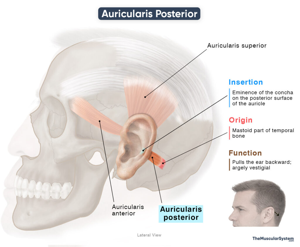
Origin
The muscle originates via short bands of aponeurosis from the surface of the mastoid part of the temporal bone, just behind the auricle or external ear.
Insertion
The originating fibers course medially as they form a thicker muscle belly. Unlike the other two auricular muscles, the auricularis posterior belly consists of two or three narrow fascicles. The muscle fibers course towards the back of the ear and insert into the eminence of the concha at the lower back aspect of the cranial surface of the auricle.
Relations With Surrounding Muscles and Structures
As evident from the name, the auricularis posterior is the most posterior of the three auricular muscles. It is also the smallest of the three, lying superficial to the deep temporal fascia, and it overlies the posterior auricular artery and vein, which ascend deep to the muscle before supplying the auricle.
Function
| Action | Pulling the ear backward |
Similar to the other auricular muscles, this muscle contracts to draw the auricle backward. Although it no longer produces meaningful movement in humans, it once played a role in orienting the ears toward sounds in early primates. Evidence indicates that the muscle still activates when we try to listen intently. In people who can “wiggle” their ears, the muscle’s movements may be more voluntary.
Recent cadaveric studies show that the muscle consistently overlies the sigmoid sinus and the transverse–sigmoid junction, making it a useful superficial landmark for neurosurgical procedures in the posterior fossa, where the cerebellum is located.
Antagonists
As with the other auricular muscles, it does not require a direct antagonist because its movements are minimal. Once the muscle relaxes, the auricle simply returns to its resting position on its own.
Innervation
| Nerve | Posterior auricular nerve |
The muscle is innervated by the posterior auricular branch of the facial nerve (CN VII).
Blood Supply
| Artery | Posterior auricular artery |
It receives its primary blood supply from the posterior auricular artery, which is a branch of the external carotid artery.
References
- Auricularis Posterior Muscle: Elsevier.com
- Auricularis Posterior Muscle: IMAIOS.com
- Posterior Auricular Muscle: Radiopaedia.org
Della Barnes, an MS Anatomy graduate, blends medical research with accessible writing, simplifying complex anatomy for a better understanding and appreciation of human anatomy.
- Latest Posts by Della Barnes, MS Anatomy
-
Tensor Tympani
- -
Stapedius
- -
Auricularis Superior
- All Posts


