

Cricothyroid
By
Della Barnes, an MS Anatomy graduate, blends medical research with accessible writing, simplifying complex anatomy for a better understanding and appreciation of human anatomy.
Last updated:
05/01/2026Della Barnes, MS Anatomy
UX/UI Designer at - AdobeDella Barnes, an MS Anatomy graduate, blends medical research with accessible writing, simplifying complex anatomy for a better understanding and appreciation of human anatomy.
The cricothyroid is a small paired muscle located deep in the lower neck region, at the front of the larynx. It is one of the intrinsic muscles of the larynx, with the others being the posterior cricoarytenoid, lateral cricoarytenoid, oblique arytenoid, transverse arytenoid, and thyroarytenoid.
It is one of the muscles of vocalization, mainly helping with changing the tone of the voice by tensing the vocal cords, earning the name, singer’s muscle.
Anatomy
Location and Attachments
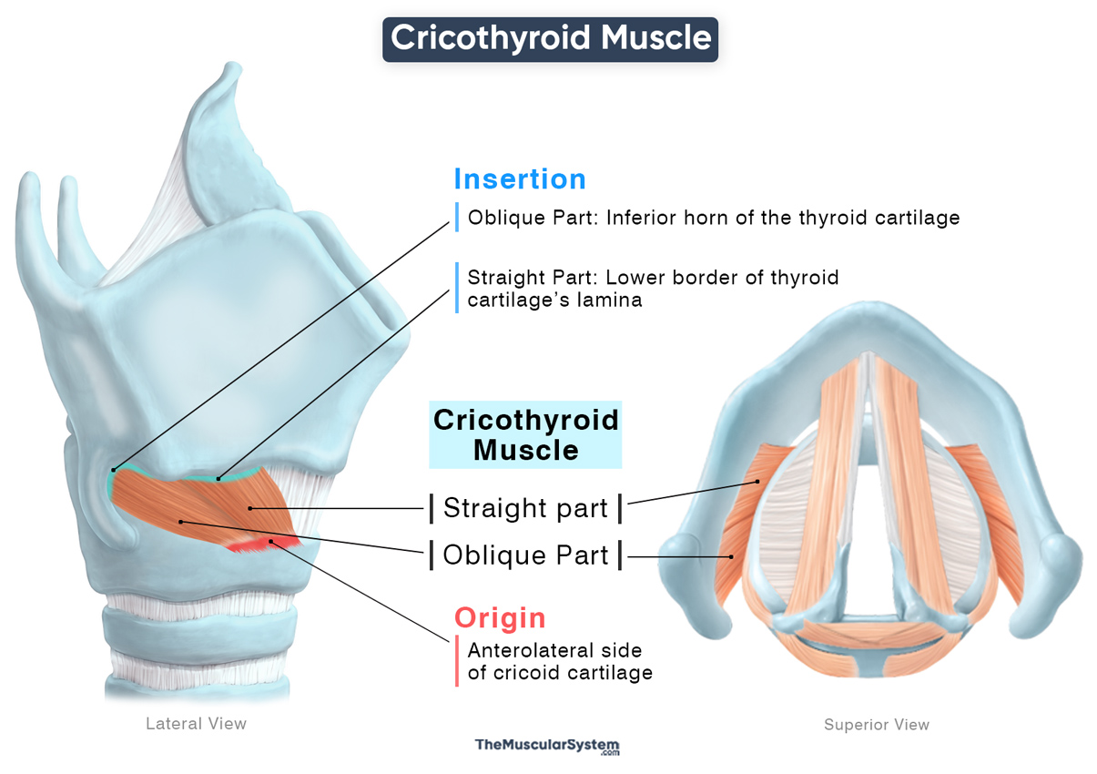
| Origin | Anterolateral aspect of the cricoid cartilage |
| Insertion | Straight Part: Inferior border of the lamina of the thyroid cartilage Oblique Part: Inferior cornu (horn) of the thyroid cartilage |
Origin
The muscle lies between the cricoid and thyroid cartilages, originating via a narrow point from the anterolateral surface of the cricoid cartilage, near the cricoid arch.
Insertion
From the point of origin, the muscle fibers fan out and separate into two groups, forming the two parts of the muscle, named for the orientation of their fibers:
Straight Part: It is the upper portion of the muscle that courses posteriorly, almost straight upward, inserting into the sides of the lower border of the thyroid cartilage’s lamina.
Oblique Part: It is the lower part of the muscle that courses obliquely and laterally towards the back, and then inserts into the inferior cornu of the thyroid cartilage.
Relations With Surrounding Muscles and Structures
The muscle lies above and superficial to the lateral cricoarytenoid muscle, and deep to the inferior thyroid contrictor, specifically near the origin of its thyropharyngeal part. Since it lies beneath the thyroid cartilage, it is also deep to the infrahyoid muscles.
It is slightly behind the inferior margin of the thyroid cartilage, and medial to the joint between the cricoid and thyroid cartilages (the cricothyroid joint). The left and right lobes of the thyroid gland partially overlay the muscles. All these relations make the cricothyroid an important anatomical and medical landmark.
Due to its close proximity to these cartilages, the muscle may sometimes suffer injury due to cricothyroidotomy. It is an emergency procedure in which a temporary airway is created through the cricothyroid membrane to allow breathing when other methods fail.
Function
| Action | Tensing the vocal folds to help with high-pitched vocalization |
The cricothyroid is the primary laryngeal tensor muscle that works on the vocal folds, playing a key role in pitch modulation during vocalization. Here is a step-by-step sequence of how it works:
- When the muscle contracts, it pulls on its insertion at the thyroid cartilage, rotating the thyroid cartilage forward and downward at the cricothyroid joint, while the cricoid cartilage remains relatively fixed at the origin.
- This movement increases the distance between the thyroid cartilage and the arytenoid cartilage — a paired laryngeal cartilage located behind the thyroid cartilage.
- Since the vocal cords attach to the thyroid cartilage in front and to the arytenoid cartilages behind, the increased distance between the two makes the vocal cords stretch and become tighter.
- Increased vocal fold tension allows them to vibrate at a higher frequency, producing higher-pitched sounds during speech and singing. to vibrate at a higher frequency, producing higher-pitched sounds during speech and singing.
Though this muscle is involved in raising the pitch of the voice, it is not the same as increasing the volume. That depends mainly on airflow and subglottic pressure.
Antagonists
The thyroarytenoid muscle can be considered antagonistic to this muscle as it decreases tension on the vocal cords by shortening and relaxing them, thereby reducing pitch during vocalization.
Innervation
| Nerve | External laryngeal nerve |
The cricothyroid muscle is innervated by the external laryngeal nerve, arising from the superior laryngeal nerve of the vagus nerve (CN X), which reaches the muscle from above. It is the only intrinsic laryngeal muscle supplied by the superior laryngeal nerve, as the rest are all innervated by the recurrent laryngeal nerve (CN X).
Blood Supply
| Artery | Cricothyroid artery |
The muscle is supplied by the cricothyroid artery, a branch of the superior thyroid artery from the external carotid artery.
References
- Cricothyroid Muscle: Kenhub.com
- Cricothyroid Muscle (Left): Elsevier.com
- Cricothyroid: TeachMeAnatomy.info
- Cricothyroid Muscle: IMAIOS.com#
Della Barnes, an MS Anatomy graduate, blends medical research with accessible writing, simplifying complex anatomy for a better understanding and appreciation of human anatomy.
- Latest Posts by Della Barnes, MS Anatomy
-
Tensor Tympani
- -
Stapedius
- -
Auricularis Posterior
- All Posts


