

Trapezius
By
Della Barnes, an MS Anatomy graduate, blends medical research with accessible writing, simplifying complex anatomy for a better understanding and appreciation of human anatomy.
Last updated:
11/07/2023Della Barnes, MS Anatomy
UX/UI Designer at - AdobeDella Barnes, an MS Anatomy graduate, blends medical research with accessible writing, simplifying complex anatomy for a better understanding and appreciation of human anatomy.
What is the Trapezius
The trapezius is a large muscle extending from the base of the neck down to the upper-middle back. It is a superficial muscle and an extrinsic muscle of the back. Still, it is considered an upper limb muscle instead of a back muscle because of its involvement in the movement of the shoulder girdle. The muscle also plays a vital role in all neck movements and in keeping your spine straight.
The human body has two trapezius muscles, each with a distinct triangular shape. As the left and right trapezius are located side by side, they resemble a trapezoid or a diamond shape, hence the name.
Anatomy
Location and Attachments
The trapezius consists of long muscle fibers that span a large area between the upper back, neck, and shoulders, ending at a single point in the mid-back. It is one of the broadest muscles in the upper back of the human body, and its fibers divide into the superior (descending), middle (transverse), and inferior (ascending) portions.
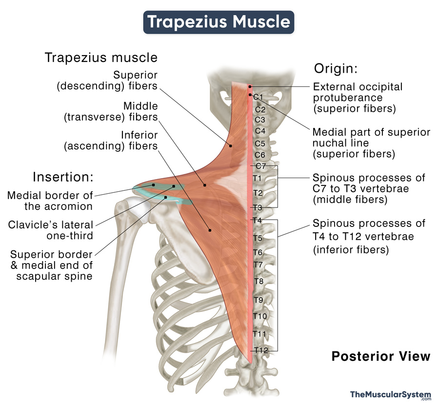
| Origin | Superior fibers: Medial part of the superior nuchal line and the external occipital protuberance Middle fibers: Superior aspect of the spinous processes of the C7 to T3 vertebrae Inferior fibers: Inferior aspect of the spinous processes of the T4 to T12 |
| Insertion | Superior fibers: Posterior border of the clavicle’s lateral one-third Middle fibers: Medial border of the scapula’s acromion, and superior border of the scapular spine Inferior fibers: Medial end of the spine of the scapula |
Origin
The trapezius muscle has a broad origin from the superior nuchal line (on the occipital bone) and the external occipital protuberance to the spinous processes of the 7th cervical and all the thoracic vertebrae. A part of the superior fibers arises from the nuchal ligament, which extends over the 1st to 7th cervical vertebrae. The supraspinal ligament is another minor point of origin for all three muscle parts.
Insertion
After originating, the upper or superior portion of the muscle course downwards laterally to reach its insertion point into the lateral one-third of the clavicle’s posterior border. Similarly, the middle portion courses horizontally and laterally to insert into the scapula – at the medial border of its acromion and the scapular spine’s upper border. The lower or inferior portion courses upwards laterally and converge into an aponeurosis near the scapula. It then passes over the scapular spine to insert into a tubercle at the lateral apex of its medial end.
The direction of the muscle fibers of the superior, middle, and inferior portions gain them their respective names of descending, transverse, and ascending parts of the trapezius.
Relations With Surrounding Muscles and Structures
As mentioned above, the muscle has a diamond shape, with the two side angles marking the two shoulders, the top angle corresponding to the occipital protuberance, and the lower angle to the spinous process of T12.
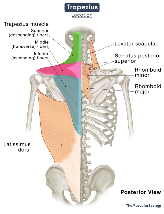
It forms the superficial layer of extrinsic back muscles along with the latissimus dorsi, rhomboid major, rhomboid minor, and levator scapulae. The trapezius is the most superficial of these muscles, with the other 4 superficial extrinsic back muscles lying deep. Additionally, the serratus posterior superior, from the intermediate layer of extrinsic back muscles, lies deep to the trapezius.
Since it is a broad muscle, the trapezius also covers several superficial intrinsic back muscles, including the iliocostalis, longissimus, spinalis, splenius cervicis, and splenius capitis. The muscle’s superior fibers overlie the muscles in the suboccipital region (rectus capitis posterior major and minor and obliquus capitis superior and inferior). The trapezius and the semispinalis capitis muscle in the neck form the primary part of the muscular mass in the occipital region at the skull’s base.
The anterior border of this muscle forms the posterior margin of the posterior neck triangle. The edges of its inferior portion make up the medial margins of the triangle of auscultation on both sides. The triangle of auscultation is a small anatomical region on the back that is only covered by a thin layer of muscle instead of being hidden under the scapula. It helps with a better pulmonary examination.
Function
| Action | Laterally rotating, elevating, and retracting the scapula; extending and laterally flexing the neck when the scapula is fixed. |
The three portions of the trapezius stabilize the scapula during shoulder and arm movements and help move the neck.
Superior (Descending) Portion
Leveling the scapula: This part of the muscle works with the levator scapulae to elevate the scapula along the scapulothoracic joint, like when you shrug your shoulders. Similarly, the muscle stabilizes the scapula to brace the shoulders against gravity when you carry something heavy in your hands.
Flexing the neck: When the muscle’s left or right side acts alone (unilaterally), it flexes the head and neck at the corresponding atlanto-occipital joint so you can bend your head to that side.
Rotating the neck: When the superior fibers contract unilaterally, it also acts on the atlantoaxial joint, allowing the head to rotate from side to side.
Extending the head and neck: When both sides of the muscle’s superior fibers work together (bilateral contraction), it helps extend the head and neck when other muscles keep the scapula stable in its place. The trapezius brings the occipital bone closer to the scapula in this action.
Middle (Transverse) Portion
Adducting and retracting the scapula: The transverse fibers work with the rhomboids to adduct the scapula, spreading them outwards and then retracting or pulling them back towards the midline.
Inferior (Ascending) Portion
Lowering the shoulders: The inferior fibers help lower the shoulders by depressing scapula medially, bringing them closer to the inferior part of the thoracic vertebrae. This action helps lower the shoulder against upward resistance, like when you push yourself up with your hands to stand up from a seating position.
Rotating the scapula: The inferior portion works together with the superior portion to laterally push the scapula’s inferior angle, raising the acromion and producing a rotating movement.
General Functions Involving the Whole Muscle
Helping raise the arm: The trapezius and serratus anterior work synergistically to rotate the scapula upwards so you can raise your arms over the should level. It means the trapezius anchors the scapula in place, so the scapulohumeral muscles (the muscles that attach the scapula to the humerus and upper arm) can function. So, you would not be able to use your arms without the trapezius.
The trapezius muscle prevents unnecessary scapula movements and keeps it stable by contracting all its fibers simultaneously.
The serratus anterior, latissimus dorsi, and pectoralis major muscles act as the antagonists of the trapezius.
Innervation
| Nerve | Anterior rami of spinal nerves (C3 and C4); accessory nerve |
It is the only upper limb muscle not to be innervated by the brachial plexus. It receives motor innervation from the anterior rami of spinal nerves C3 and C4 and the accessory nerve, also known as the cranial nerve XI (CN XI). The C3 and C4 spinal nerves also contain the muscle’s sensory fibers that help with proprioception and sensations like pain.
Blood Supply
| Artery | Transverse cervical artery |
The primary blood supply comes from the transverse cervical artery, with the dorsal scapular artery supplying the superior portion. The posterior intercostal arterial branches provide the deep parts of the muscle.
The dorsal scapular artery forms a more significant part of the muscle’s blood supply in an anatomical variation.
References
- Trapezius Muscle: InnerBody.com
- Trapezius: TeachMeAnatomy.info
- Trapezius Muscle: KenHub.com
- Anatomy, Back, Trapezius: NCBI.NLM.NIH.gov
- Trapezius Muscle: Clevelandclinic.org
- Trapezius Muscle: GetBodySmart.com
- Trapezius: Meddean.LUC.edu
- Trapezius: Action, Origin, Insertion, & Attachments: Study.com
Della Barnes, an MS Anatomy graduate, blends medical research with accessible writing, simplifying complex anatomy for a better understanding and appreciation of human anatomy.
- Latest Posts by Della Barnes, MS Anatomy
-
Tensor Tympani
- -
Stapedius
- -
Auricularis Posterior
- All Posts


