

Latissimus Dorsi
By
Della Barnes, an MS Anatomy graduate, blends medical research with accessible writing, simplifying complex anatomy for a better understanding and appreciation of human anatomy.
Last updated:
23/05/2025Della Barnes, MS Anatomy
UX/UI Designer at - AdobeDella Barnes, an MS Anatomy graduate, blends medical research with accessible writing, simplifying complex anatomy for a better understanding and appreciation of human anatomy.
What is Latissimus Dorsi
Latissimus dorsi, often known as the ‘lats,’ is the broadest muscle in the human body that covers almost the entire back, overlying most other back muscles. The flat spine muscle is one of the four superficial extrinsic back muscles, along with the trapezius, rhomboids, and levator scapulae.
It plays a vital role in arm movement and also helps with respiration.
Anatomy
Location and Attachments
It is a paired muscle that spreads over a large area, covering the lower thoracic vertebrae and all the lumbar vertebrae (middle and lower back) and extending to the lateral sides of the armpits. The left and right latissimus dorsi cover the left and right sides of the back and can be partially visible from the front when the arms are raised.
The muscle, with its multiple points of attachment, gives the torso its ‘V’ shape.
| Origin | Vertebral attachment: The spinous processes of the T7 to T12 vertebrae; The thoracolumbar fascia Iliac attachment: Posterior 1/3rd of the iliac crest; Costal attachment: The lower 3 or 4 ribs (9th to 12th) Scapular attachment: The scapula’s inferior angle |
| Insertion | The bicipital groove (Intertubercular sulcus) on the anterior side of the humerus |
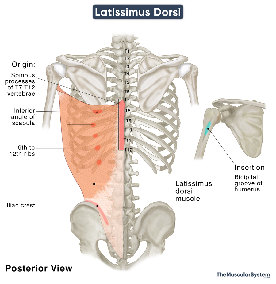
Origin
Being the broadest muscle, it has 5 different points of origin, named after the regions. Thin tendinous fibers arise from the T7 to T12 vertebrae. Some muscle fibers arise from the lumbodorsal fascia, which attaches the muscle to the lumbar and sacral vertebral spines. Another point of origin is from the posterior border of the iliac crest. At the same time, multiple fleshy strips arise from the 9th or 10th to the 12th ribs.
After originating from all these points, the muscle fibers course in different directions – the superior fibers run horizontally, the middle fibers run obliquely, and the inferior fibers run vertically. Together they cross the scapula, where a few muscle fibers arise from its inferior angle. The muscle fibers converge into a single fasciculus near the inferior scapula, which curves around the lower side of the Teres major muscle. The fasciculus also twists upon itself, so the superior fibers turn posterior before being positioned on the inferior side. Similarly, the inferior fibers go anterior before going into the superior position.
Insertion
The muscle ends in a 2-3 inches long tendon that inserts into the lower end of the bicipital groove of the humerus. The muscle’s insertion lies higher on the humerus than that of the Pectoralis major, while the tendon of the teres major inserts just below the lat. A bursa separates the inserting tendons of the lat and the teres major.
A simple mnemonic can help remember the order of insertion of these three muscles:
“Lady between two majors”
Lady: Latissimus dorsi
Majors: Pectoralis major (above) and teres major (below)
Relations With Surrounding Muscles and Structures
As mentioned above, it is a superficial back muscle covering the middle and lower back. The trapezius is the only muscle that lies superficial to the lats, while the serratus posterior lies deep to it.
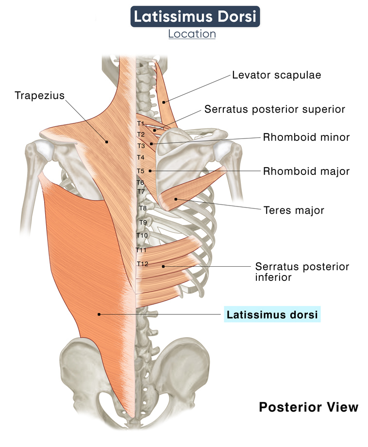
Latissimus dorsi, accompanied by the teres major, runs through the space between the scapula and the proximal part of the humerus. Here the two muscles form the inferior aspect of the posterior axillary fold, which is the lower back side of the armpit. The fold accentuates when the arm is pulled back (adducted) towards the trunk against resistance, like when climbing. When the muscle is in this position, it is possible to trace its entire inferolateral course up to its iliac attachment.
The inferolateral margin of the lats forms the medial border of the Petit’s triangle or inferior lumbar triangle, a small anatomical space in the lower back. The external and internal abdominal oblique muscles and the iliac crest further bind this space.
It also contributes to forming the inferior border of the auscultation triangle, another vital anatomical landmark in the mid-back region—the trapezius muscle and the scapula’s medial border form this space’s superior and medial borders.
Function
Its wide attachment involving multiple points of attachment and the multidirectional orientation of its muscle fibers allow the lats to help with several vital movements.
| Action | Internally rotating, adducting, and extending the arms; assisting in respiration with its costal attachment |
Helping with arm movement
This muscle plays a vital role in activities like climbing, rowing, and swimming.
Through its insertion point at the proximal humerus, the muscle works with the teres and pectoralis major to adduct, extend, and medially rotate the arm at the glenohumeral joint. When the lats contract, the back extends and rotates so the arms can extend overhead, like when reaching for something on a high wall shelf. Both adduction and extension are the strongest when the arm is already partially flexed or abducted.
Another significant action of this muscle is pulling the trunk forward and upward when the humerus remains fixed in the glenoid cavity of the scapula. During these movements, the lats work in synergy with the pectoralis major. It helps with climbing and doing exercises like pull-ups as well as assists people who walk with crutches to pull their bodies forward.
During movements of the humerus and the shoulder joint, the latissimus dorsi holds the scapula stable against the thoracic cage at the scapulothoracic joint.
Assisting respiration
Since the muscle is attached to the spinous processes of multiple thoracic vertebrae, it assists respiration as an accessory respiratory muscle. Studies have shown it to help expand the rib cage during deep inhalation or inhalation against resistance. Similarly, it assists with compressing the rib cage during forced expiration, like with coughing or sneezing.
Antagonists
The latissimus dorsi has several muscles that act as antagonists because it acts on a vast region on the shoulder, arms, thoracic cage, and back. Its primary antagonists include the biceps brachii, coracobrachialis, deltoids, supraspinatus, infraspinatus, teres minor, and pectoralis major.
Innervation
| Nerve | Thoracodorsal nerve (C6 to C8) |
Its innervation comes from the thoracodorsal nerve arising from the posterior cord of the brachial plexus from nerve roots C6, C7, and C8.
Blood Supply
| Artery | Thoracodorsal artery |
Blood supply to this muscle comes from the thoracodorsal artery. It is a branch of the subscapular artery that enters from the costal surface of the muscle, with only a few inches between it and the subscapular artery.
Additional blood supply comes from the perforating branches of the 9th to 11th posterior intercostal arteries and the 1st to 3rd lumbar arteries.
References
- Anatomy, Back, Latissimus Dorsi: NCBI.NLM.NIH.gov
- Latissimus Dorsi: Structure and Function: Study.com
- Latissimus Dorsi Muscle: IMAIOS.com
- Latissimus Dorsi Muscle: KenHub.com
- Latissimus Dorsi Muscle – Attachments, Action & Innervation: GetBodySmart.com
- Latissimus Dorsi: TeachMeAnatomy.info
- Latissimus Dorsi Muscle: RadioPaedia.org
- Latissimus Dorsi: Action, Muscles & Antagonist: Study.com
Della Barnes, an MS Anatomy graduate, blends medical research with accessible writing, simplifying complex anatomy for a better understanding and appreciation of human anatomy.
- Latest Posts by Della Barnes, MS Anatomy
-
Tensor Tympani
- -
Stapedius
- -
Auricularis Posterior
- All Posts


