

Deltoid
By
Della Barnes, an MS Anatomy graduate, blends medical research with accessible writing, simplifying complex anatomy for a better understanding and appreciation of human anatomy.
Last updated:
23/04/2024Della Barnes, MS Anatomy
UX/UI Designer at - AdobeDella Barnes, an MS Anatomy graduate, blends medical research with accessible writing, simplifying complex anatomy for a better understanding and appreciation of human anatomy.
What is the Deltoid
The deltoid is a large, thick triangular skeletal muscle in the shoulder joint, connecting the arm to the body trunk. Its name derives from ‘delta’ (Δ), the Greek letter that it resembles in shape.
The deltoids give our shoulder its rounded shape and are responsible for most major arm movements.
Anatomy
Location and Attachments
Deltoids or delts lie over each arm’s glenohumeral or shoulder joint and are the most superficial muscle in this area. It means that the deltoids are located closest to the skin surface of all the shoulder muscles.
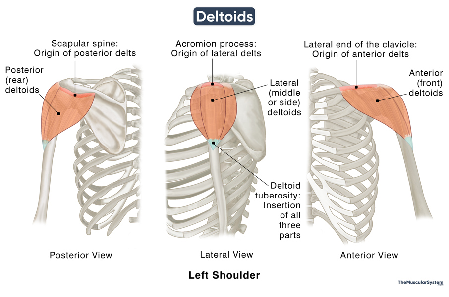
The muscle is the widest at the top, formed of three distinct sets of muscle fibers, which divide it into three parts or ‘heads’ with different points of origin.
| Origin | Anterior (front) deltoids: One-third of the lateral end of clavicle Lateral (middle or side) deltoids: The upper surface and lateral margin of the acromion of scapula Posterior (rear) deltoids: Lateral one-third of the scapular spine |
| Insertion | Deltoid tuberosity of the humerus |
As the muscle travels down the shoulder and upper arm, the three parts come together into one narrow solid tendon that attaches to the deltoid tuberosity around the middle of the anterior humeral shaft.
Relations to Other Muscular Structures
Since it is a large muscle located superficially, it overlies several other muscles in the shoulder and upper arm. These include 4 rotator cuff muscles: supraspinatus, infraspinatus, subscapularis, and teres minor. The pectoralis major, tendons of pectoralis minor, and coracobrachialis, the two heads of biceps, and the lateral head of triceps are also covered by the deltoids.
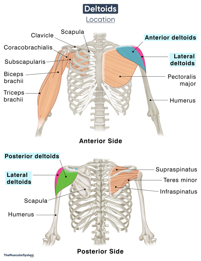
Apart from the above muscular structures, the delts cover the subacromial bursa, coracoacromial ligament, coracoid process of the scapula, the proximal part of the humerus, the axillary nerve, as well as the posterior circumflex humeral artery and nerve.
Functions
| Action | Anterior delts flex and medially rotate the arm; Lateral delts abduct the arm; Posterior delts extend and laterally rotate the arm; |
The deltoids are the biggest and primary muscles in the shoulder region, being the main abductors of the arm at the shoulder joint. They allow you to move your arms in almost any direction. However, for the delts to take over the abduction, the arm must already be abducted by about 15 degrees. The supraspinatus muscle takes care of this initial abduction.
The pectoralis major is the primary antagonist of the deltoid on the anterior side. In contrast, on the posterior side, it’s the latissimus dorsi.
They keep the shoulder joint in place, contracting and relaxing the shoulders during all arm movements. The three parts divide different actions and movements among themselves.
Anterior deltoids: Helps flex the arms forward in front of the body, for example, when reaching out to pick something up or get something from a shelf. It also helps in rotating the arm medially, like when you cross your arms or reach across your own body.
Lateral deltoids: The largest, middle part of the delts helps you move your arms out on your sides. This is the muscle you would use to raise your arms as you do jumping jacks.
Posterior deltoids: Helps extend the arm backward, for example, when you pitch a baseball or practice arm circling. This is the smallest of the three parts and is used the least.
Innervation
| Nerve | Axillary nerve (C5 and C6) |
Blood Supply
| Artery | Thoracoacromial artery, subscapular artery, anterior and posterior circumflex humeral artery, and deep brachial artery |
Being such a large muscle, the deltoid requires several arteries to supply it. All the arteries are axillary artery branches except the deep brachial artery, which, as the name suggests, is a branch of the brachial artery. It is itself a branch of the axillary artery and the primary source of arterial supply in the arm.
References
- Deltoid Muscles: ClevelandClinic.org
- Deltoid muscle: KenHub.com
- Deltoid: Rad.Washington.edu
- Deltoid: TeachMeAnatomy.info
- What Is the Deltoid Muscle: Study.com
- Deltoid Muscle: InnerBody.com
- Deltoid Muscle – Anterior and Middle Heads: GetBodySmart.com
Della Barnes, an MS Anatomy graduate, blends medical research with accessible writing, simplifying complex anatomy for a better understanding and appreciation of human anatomy.
- Latest Posts by Della Barnes, MS Anatomy
-
Tensor Tympani
- -
Stapedius
- -
Auricularis Posterior
- All Posts


