

Pectineus
By
Della Barnes, an MS Anatomy graduate, blends medical research with accessible writing, simplifying complex anatomy for a better understanding and appreciation of human anatomy.
Last updated:
19/08/2025Della Barnes, MS Anatomy
UX/UI Designer at - AdobeDella Barnes, an MS Anatomy graduate, blends medical research with accessible writing, simplifying complex anatomy for a better understanding and appreciation of human anatomy.
What is the Pectineus
The pectineus is a short, flat, quadrangular muscle located in the upper front of the thigh. It belongs to the medial compartment of the hip, along with the gracilis, adductor brevis, adductor longus, and adductor magnus.
The small muscle plays a key role in the adduction and flexion of the thigh at the hip joint. These movements are essential for coordinated lower limb function during activities such as walking, climbing stairs, and maintaining posture.
Anatomy
Location and Attachments
| Origin | Pectineal line of the pubis |
| Insertion | Pectineal line of the femur |
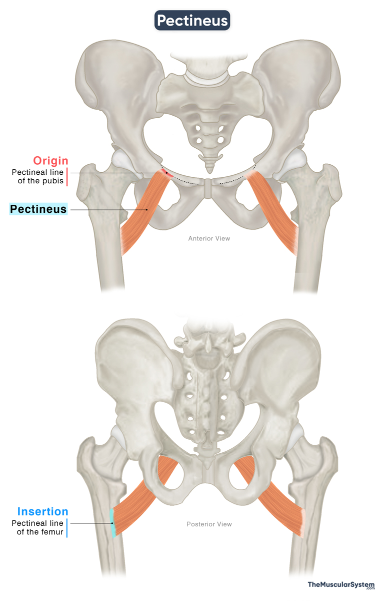
Origin
The muscle originates via short tendons from the pecten pubis or pectineal line of the pubis bone and the adjacent bony surface and fascial layers.
Insertion
From its origin, the muscle belly descends laterally, slightly towards the back, to cross the hip joint capsule on its medial side. Then it inserts into the pectineal line of the femur, a small ridge located between the lesser trochanter and the linea aspera.
Relations With Surrounding Muscles and Structures
The pectineus is the most anteriorly positioned hip adductor and has the highest point of origin among the muscles in this group. It lies just deep to the fascia lata, which is the fibrous layer of connective tissue that surrounds the thigh muscles. The obturator foramen is situated behind and is covered by its quadrangular muscle belly.
Medially, the adductor longus lies adjacent to the pectineus, and together they form the medial portion of the floor of the femoral triangle, a space in the upper thigh that allows the passage of several nerves, blood vessels, and other structures to the lower limb. Notable structures passing through the femoral triangle and lying superficial to the pectineus include the femoral artery, femoral vein, and great saphenous vein.
Laterally, the pectineus is bordered by the psoas major, while the obturator externus, adductor brevis, and adductor magnus lie deep or posterior to it.
Function
| Action | Adducting and flexing the thigh at the hip joint |
As one of the primary hip adductors, pectineus plays a major role in both flexing and adducting the hip joint. When the muscle contracts, it first helps flex or bend the thigh at the hip joint, up to about 45°. Once the hip is flexed, further contraction of the muscle helps adduct the thigh, bringing the thighs closer together.
A movement that uses both actions of the pectineus is crossing the legs at the knee: the thigh first flexes at the hip, then moves inward toward the midline to complete the crossing.
When the muscle contracts solely to flex the hip, it acts in concert with the iliopsoas, rectus femoris, and sartorius to assist in the terminal swing phase of the gait cycle. Minor functions include contributing to hip joint stability, and some sources suggest it may also play a role in internal rotation of the hip.
Antagonists
The gluteus maximus and biceps femoris are the primary hip extensors, making them antagonistic to the pectineus as a hip flexor.
For its adduction role, the main antagonists are the gluteus medius, gluteus minimus, and tensor fasciae latae, which are the primary hip abductors.
Innervation
| Nerve | Femoral nerve (L2-L3) |
While the obturator nerve innervates most muscles of the medial thigh compartment, the pectineus is an exception. Its main nerve supply comes from the femoral nerve, specifically from the first and second lumbar nerve roots (L2-L3). Because the femoral nerve mainly supplies muscles in the anterior compartment, the pectineus is sometimes classified as part of that rather than the medial compartment.
It may also receive a secondary contribution from the obturator nerve (L2–L3).
Blood Supply
| Artery | Medial circumflex femoral artery, obturator artery |
The medial circumflex femoral artery supplies the anterior or superficial portion of the muscle. This artery branches from the femoral artery, which arises from the external iliac artery.
The posterior or deep part of the muscle gets its blood supply from the anterior branch of the obturator artery, a branch of the internal iliac artery.
References
- Pectineus: Osmosis.org
- Pectineus: TeachMeAnatomy.info
- Pectineus Muscle: Elsevier.com
- Pectineus Muscle: Radiopaedia.org
- Pectineus Muscle: Kenhub.com
- Anatomy, Bony Pelvis and Lower Limb: Medial Thigh Muscles: NCBI.NLM.NIH.gov
- Pectineus Muscle: GetBodySmart.com
Della Barnes, an MS Anatomy graduate, blends medical research with accessible writing, simplifying complex anatomy for a better understanding and appreciation of human anatomy.
- Latest Posts by Della Barnes, MS Anatomy
-
Tensor Tympani
- -
Stapedius
- -
Auricularis Posterior
- All Posts


