

Adductor Magnus
By
Della Barnes, an MS Anatomy graduate, blends medical research with accessible writing, simplifying complex anatomy for a better understanding and appreciation of human anatomy.
Last updated:
22/08/2025Della Barnes, MS Anatomy
UX/UI Designer at - AdobeDella Barnes, an MS Anatomy graduate, blends medical research with accessible writing, simplifying complex anatomy for a better understanding and appreciation of human anatomy.
What is the Adductor Magnus
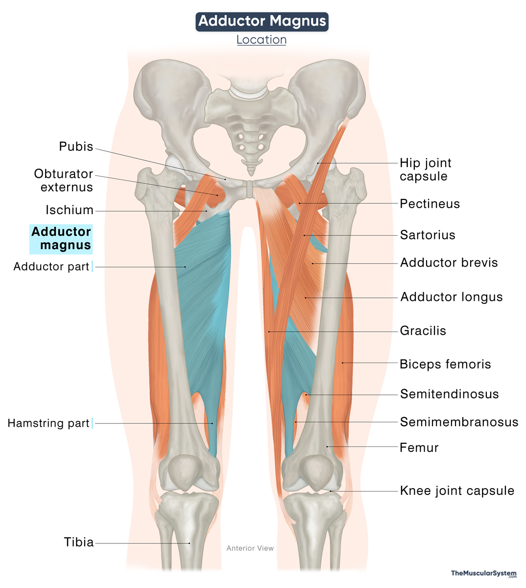
The adductor magnus is a large triangular muscle that extends along the inner thigh region. It belongs to the group of hip adductors alongside the gracilis, pectineus, adductor brevis, and adductor longus. It is one of the primary muscles for the movement of the lower limb.
It is the largest and strongest muscle in the medial compartment of the thigh, and has two parts, the adductor portion and the hamstring portion. Though typically considered to be a part of the medial compartment, the hamstring portion of the muscle is sometimes included in the posterior compartment.
Anatomy
Location and Attachments
The two parts of the muscle have separate points of origin and insertion, and are named accordingly. The shorter, adductor part is also known as the pubofemoral part, while the hamstring part is also referred to as the ischiocondylar part.
| Origin | Adductor (pubofemoral) part: Inferior pubic ramus and ischial ramus Hamstring (ischiocondylar) part: Ischial tuberosity |
| Insertion | Adductor part: Gluteal tuberosity, medial lip of the linea aspera, and the medial supracondylar line of the femur Hamstring part: Medial supracondylar line, and adductor tubercle of the femur |
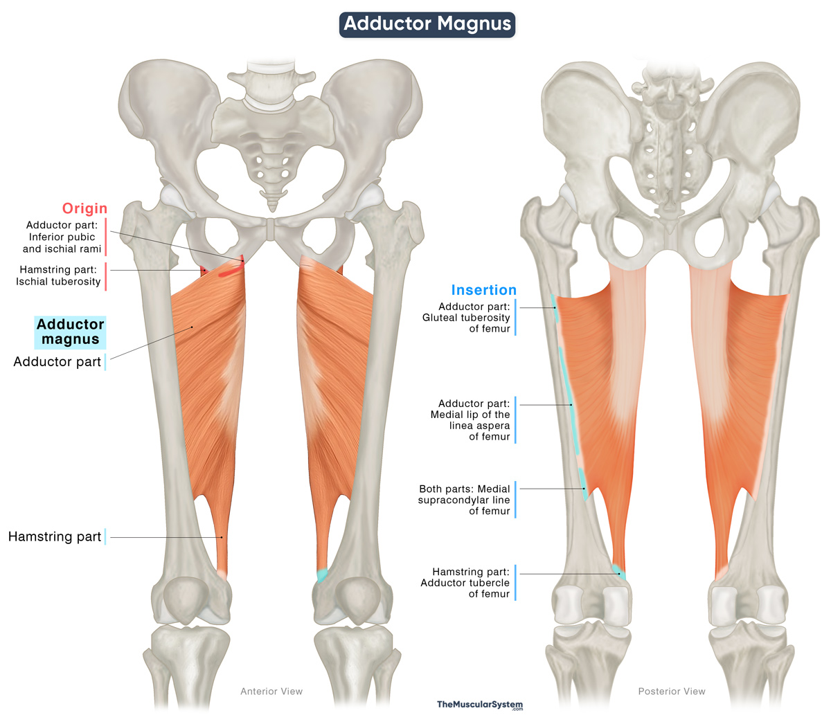
Adductor (Pubofemoral) Part
Origin
The pubofemoral part of the muscle originates from two points, with the orientation of the muscle fibers varying based on the origin. The superior (upper) segment originates from the ischiopubic ramus, a small bony surface where the inferior pubic ramus and the ischial ramus meet. The inferior (lower) segment rises solely from the ischial ramus.
Insertion
The superior segment of the adductor magnus runs almost horizontally from its origin to insert into the gluteal tuberosity and the upper medial part of the linea aspera, just medial to the insertion of the gluteus maximus. This portion lies more anteriorly than the rest of the muscle, which is why some sources describe the pubofemoral part as a separate muscle, referred to as the adductor minimus.
The inferior segment is considerably larger, with fibers spreading out into a broad, fan-shaped belly. These fibers course laterally and downward, converging into a wide aponeurosis that inserts along the medial lip of the linea aspera, including the upper medial supracondylar line. Here, its tendinous insertions merge with the origins of the short head of the biceps femoris along the linea aspera.
Hamstring (Ischiocondylar) Part
Origin
This part of the muscle originates from the ischial tuberosity.
Insertion
From its origin, the tendon descends almost vertically, forming a fleshy muscle belly that tapers in the lower thigh into a rounded tendon. This tendon continues downward to insert into the medial supracondylar line and the adductor tubercle on the medial epicondyle of the femur.
This portion is known as the “hamstring” part because it shares a similar origin, general shape, and nerve supply as the hamstring muscles of the posterior thigh. However, since it inserts into the femur rather than crossing the knee joint, it is not considered a true hamstring.
Structure of the Inserting Aponeurosis
The aponeurotic sheet via which the muscle inserts into the femur has five openings. The four superior openings are smaller and allow the perforating and terminal branches of the deep femoral artery to pass into the posterior compartment of the thigh.
The fifth and largest opening is the adductor hiatus — the distal end of the adductor canal, a tunnel in the mid-thigh region that carries the neurovascular structures of the lower limb. It lets the femoral vessels pass to the popliteal fossa, the diamond-shaped depression at the back of the knee.
Relations With Surrounding Muscles and Structures
Being the largest muscle in the medial thigh, the adductor magnus is related to several muscles and neurovascular structures in the area.
Muscles and Muscular Structures
Anteriorly, it is deep to the pectineus, adductor longus, and adductor brevis. Behind it lie the gluteus maximus, semimembranosus, and semitendinosus muscles. Along its medial border run the gracilis and sartorius, while the obturator externus and quadratus femoris are superior to it.
The adductor magnus and adductor longus form the posterior wall of the adductor canal, or Hunter’s canal.
Neurovascular Structures
The femoral and deep femoral arteries and veins, along with the posterior branches of the obturator artery, nerve, and vein, are located anterior to the muscle. Posterior to it lies the sciatic nerve, which is the largest nerve in the body.
Function
| Action | — Adducting the thigh at the hip joint (both parts) — Flexing the thigh (adductor part) — Extending the thigh (hamstring part) |
Both parts in synergy
- Thigh adduction: As the strongest muscle in the medial thigh, the adductor and hamstring parts work together to adduct the thigh. It is most active when the thigh is abducted, drawing the legs back toward the midline. The muscle also plays a key role in actions such as kicking a ball with the inner side of the foot.
- Hip stabilization: Being strong adductors, both parts participate in the gait cycle, stabilizing the pelvis to control proper posture.
Adductor part
- Thigh flexion: The adductor part, particularly its superior portion with horizontally aligned fibers, assists in flexing the thigh.
- Thigh lateral rotation: With oblique fibers inserting along the linea aspera, this part also contributes, though weakly, to rotating the thigh laterally or outward.
Hamstring part
- Thigh extension: The hamstring part assists in extending the thigh, an action opposite to the flexion performed by the adductor part.
- Thigh medial rotation: With its long fibers attaching as low as the medial epicondyle, this part of the muscle weakly assists in rotating the thigh medially or inward.
Antagonists
Since it is the strongest hip adductor, its primary antagonists are the hip abductor muscles, namely the gluteus medius, gluteus minimus, and tensor fasciae latae.
Innervation
| Nerve | Adductor part: Posterior branch of obturator nerve (L2-L4) Hamstring part: Tibial branch of the Sciatic nerve (L4) |
Each part of the muscle receives innervation from a different nerve, reflecting its position across both the medial and posterior compartments of the thigh. The adductor part is innervated by the posterior branch of the obturator nerve, which arises from the second to fourth lumbar nerve roots (L2–L4).
Innervation to the hamstring part of the muscle comes from the tibial nerve, which arises from the fourth lumbar nerve root (L4). It is one of the two primary branches of the sciatic nerve.
Blood Supply
| Artery | Perforating branches of deep femoral artery, medial circumflex femoral artery, and the femoral, popliteal, and genicular arteries |
The perforating branches of the deep femoral artery are the main source of blood supply. The medial circumflex femoral artery supplies the upper portion of the muscle, while additional blood comes from the femoral, popliteal, and genicular arteries, all of which are direct or indirect branches of the deep femoral artery.
References
- Anatomy, Bony Pelvis and Lower Limb: Thigh Adductor Magnus Muscle: NCBI.NLM.NIH.gov
- Adductor Magnus: TeachMeAnatomy.info
- Adductor Magnus Muscle: Radiopaedia.org
- Adductor Magnus: Elsevier.com
- Adductor Magnus Muscle: Kenhub.com
- Adductor Magnus: Rad.UW.edu
- Adductor Magnus: IMAIOS.com
Della Barnes, an MS Anatomy graduate, blends medical research with accessible writing, simplifying complex anatomy for a better understanding and appreciation of human anatomy.
- Latest Posts by Della Barnes, MS Anatomy
-
Tensor Tympani
- -
Stapedius
- -
Auricularis Posterior
- All Posts


