

Semitendinosus
By
Della Barnes, an MS Anatomy graduate, blends medical research with accessible writing, simplifying complex anatomy for a better understanding and appreciation of human anatomy.
Last updated:
06/08/2025Della Barnes, MS Anatomy
UX/UI Designer at - AdobeDella Barnes, an MS Anatomy graduate, blends medical research with accessible writing, simplifying complex anatomy for a better understanding and appreciation of human anatomy.
What is the Semitendinosus
The semitendinosus is a thin, long, fusiform muscle located at the back of the thigh in the human body. It belongs to the group of hamstring muscles along with the biceps femoris and the semimembranosus. These three muscles also form the posterior compartment of the thigh.
Being a hamstring muscle, its primary function involves flexing the knee. The muscle gets its name from its characteristic long tendon, which comprises almost half of the muscle’s total length. You can even feel the tendon at the inner back of the knee when you bend your knee with effort, like when doing a squat.
Anatomy
Location and Attachments
| Origin | Ischial tuberosity |
| Insertion | Medial surface of the proximal tibia (via the pes anserinus) |
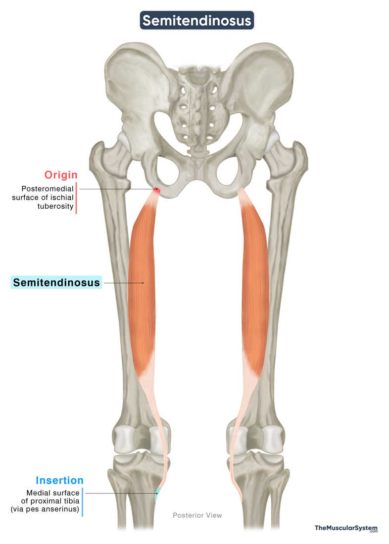
Origin
The muscle originates from the posteromedial surface of the upper ischial tuberosity. It shares this origin with the long head of the biceps femoris.
In addition, part of the semitendinosus arises from the medial side of the proximal tendon of the long head of the biceps femoris. A few muscle fibers also originate from the adjacent aponeurosis.
Insertion
The muscle fibers form a belly that runs downward along the back of the thigh. About halfway down the thigh, the belly converges into a long, narrow, rounded tendon.
As the tendon approaches the knee, it curves around the medial condyle of the tibia and continues down along the medial side of the knee joint. It passes over the medial collateral ligament, one of the major ligaments of the knee joint, and the pes anserine bursa, a small fluid-filled sac that helps reduce friction in this area. The tendon then inserts onto the anteromedial surface of the proximal tibial shaft, just below the level of the knee, as part of the pes anserinus.
Relations With Surrounding Muscles and Structures
Originating tendon and muscle belly along the thigh
At its point of origin, the semitendinosus lies deep to the originating fibers of the gluteus maximus. Within the hamstring group, it is medial to the biceps femoris and lateral to the semimembranosus. Its muscle belly and tendon travel along the posterior side of the semimembranosus and the adductor magnus, a muscle of the medial thigh compartment.
At the level of the knee, the long tendon of the muscle forms the superomedial border of the popliteal fossa, alongside the semimembranosus muscle. The popliteal fossa is a diamond-shaped depression at the back of the knee, which allows passage for several important nerves and blood vessels traveling from the thigh into the lower leg.
The insertion tendon at the tibia
At the tibia, the point of insertion of the semitendinosus tendon lies behind the inserting tendons of the gracilis and sartorius muscles. Together, these three tendons form the pes anserinus, a tendinous landmark that gets its name from the Latin for ‘goose foot,’ due to its shape resembling the webbed foot of a goose.
At this site, a bursa separates the three individual tendons, while the entire structure of the pes anserinus has the anserine bursa sitting between itself and the medial collateral ligament.
Function
| Action | Extending the thigh at the hip joint, and flexing the leg at the knee joint |
With attachments at the ischium and tibia, the semitendinosus muscle plays a vital role as part of the hamstring muscle group in the movement of both the hip and knee joints.
Which part of the lower limb it moves depends on which end of the muscle is fixed at its attachment point.
When the tibial attachment is fixed
- The muscle helps extend the thigh, like when standing up from a seated position.
- In the anatomical position, it also helps rotate the thigh inward (medial rotation).
When the ischial attachment is fixed
- It helps flex the lower leg at the knee joint, such as when you sit down in a chair.
- The muscle plays a role in medially rotating the lower leg at the knee joint, but only when the knee is already flexed beyond 90°, such as when sitting cross-legged.
Through its insertion into the medial side of the tibia, the muscle contributes to stabilizing the medial aspect of the knee joint.
Antagonists
In hip extension: The primary hip flexors, the iliopsoas and rectus femoris, act as antagonists to the semitendinosus during hip extension.
In knee flexion: The muscle’s function as a knee flexor is opposed by the quadriceps femoris group, which includes the rectus femoris, vastus lateralis, vastus intermedius, and vastus medialis, the primary extensors of the knee joint.
Innervation
| Nerve | Tibial nerve (L5-S2) |
The muscle is innervated by the tibial nerve, arising from the fifth lumbar, and the 1st and 2nd sacral nerve roots (L5, S1, S2). It is a branch of the sciatic nerve.
Blood Supply
| Artery | Inferior gluteal, and perforating arteries |
The semitendinosus muscle receives its blood supply from multiple sources. The primary vascular supply comes from the inferior gluteal artery, a branch of the internal iliac artery, as well as from the perforating arteries, which arise from the profunda femoris artery.
Additional blood supply is provided by the medial superior genicular artery, a branch of the popliteal artery.
References
- Anatomy, Bony Pelvis and Lower Limb: Thigh Semitendinosus Muscle: NCBI.NLM.NIH.gov
- Semitendinosus Muscle: Radiopaedia.org
- Semitendinosus: Rad.UW.edu
- Semitendinosus Muscle: Kenhub.com
- Semitendinosus Muscle: Elsevier.com
- Semitendinosus Muscle: IMAIOS.com
- Semitendinosus – Attachments, Actions & Innervation: GetBodySmart.com
Della Barnes, an MS Anatomy graduate, blends medical research with accessible writing, simplifying complex anatomy for a better understanding and appreciation of human anatomy.
- Latest Posts by Della Barnes, MS Anatomy
-
Tensor Tympani
- -
Stapedius
- -
Auricularis Posterior
- All Posts


