

Quadriceps
By
Della Barnes, an MS Anatomy graduate, blends medical research with accessible writing, simplifying complex anatomy for a better understanding and appreciation of human anatomy.
Last updated:
31/07/2025Della Barnes, MS Anatomy
UX/UI Designer at - AdobeDella Barnes, an MS Anatomy graduate, blends medical research with accessible writing, simplifying complex anatomy for a better understanding and appreciation of human anatomy.
What is the Quadriceps Femoris
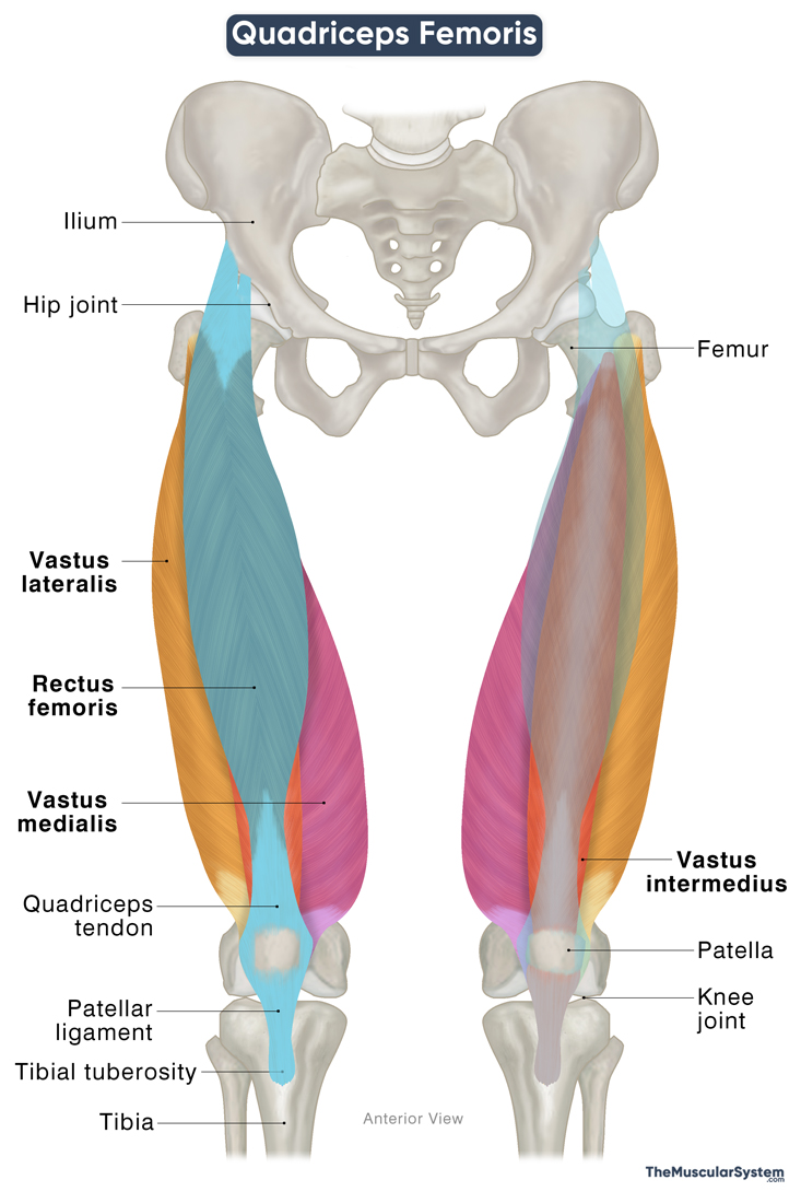
The quadriceps femoris, commonly known as the quadriceps or just “quads”, is a group of four muscles located at the front of the thigh. The name “quadriceps” comes from Latin, meaning “four-headed,” a reference to the four muscles that originate from different points but merge to form a single tendon. Together, they make up the quadriceps complex.
This is one of the strongest and largest muscle groups in the human body. With a shared attachment at the knee joint, it plays a key role in extending the knee, a movement essential for actions such as walking, running, and standing up.
The quadriceps femoris forms the anterior compartment of the thigh along with the articularis genus and sartorius muscles.
Names, Location, & Anatomy of the Muscles of the Quadriceps Group
The muscles that make up the quadriceps femoris originate from different points on the femur, except for the rectus femoris, which arises from the hip bone. The muscles get their nerve and blood supply from branches of the femoral nerve and artery.
Here is a list of the muscles:
| Name | Origin | Innervation | Blood Supply | |
|---|---|---|---|---|
| Rectus femoris | Longest and most superficial quad muscle that covers both the hip and the knee joints | — Anterior inferior iliac spine or AIIS (direct head) — Supraacetabular groove (indirect head) | Femoral nerve (L2-L4) | Descending branch of the lateral circumflex femoral artery |
| Vastus lateralis | Largest quad muscle | — Intertrochanteric line — Greater trochanter — Gluteal tuberosity — Linea aspera of the femur | Femoral nerve (L2-L4) | Transverse and descending branches of the lateral circumflex femoral artery |
| Vastus intermedius | Deepest quad muscle | Anterior surface of the proximal femoral shaft | Femoral nerve (L2-L4) | Lateral circumflex femoral artery |
| Vastus medialis | Smallest quad muscle | — Intertrochanteric line — Pectineal line — Medial lip of the linea aspera | Femoral nerve (L2-L4) | Femoral artery |
Insertion of the Common Tendon
The four quadriceps muscles descend from their respective points of origin, merging near the distal end of the femur to form the strong, broad quadriceps tendon.
This tendon inserts into the base (superior surface) of the patella (kneecap). Some of its fibers continue downward, covering the patella and forming the patellar ligament from the apex (inferior surface) of the patella. It attaches to the tibial tuberosity on the front of the proximal tibia.
While the patella serves as the initial insertion point of the quadriceps, it is actually a sesamoid bone embedded within the quadriceps tendon. Because of this, some sources describe the tibial tuberosity, where the patellar ligament attaches, as the final point of insertion. This arrangement plays a critical role in transmitting force from the quadriceps muscles to the lower leg during knee extension.
Recent Findings About a Fifth Quad Muscle
Recent anatomical studies on cadavers have identified a fifth muscle in the quadriceps complex, termed the tensor of vastus intermedius (TVI). This newly described muscle originates from the proximal femur, specifically the anterior aspect of the greater trochanter, and lies between the vastus lateralis and vastus intermedius.
Like the other quadriceps muscles, the tensor of the vastus intermedius contributes to the quadriceps tendon, which inserts into the superior surface of the patella. While its role remains under investigation, this muscle may increase the structural complexity of the quadriceps and have an impact on knee function.
Function
With attachments both at the hip joint (via rectus femoris) and the knee joint, the functions of the muscle include:
1. Extending the Knee Joint
The quadriceps femoris is the strongest and primary extensor of the leg at the knee joint. The quadriceps tendon and the patellar ligament connect the quads to the tibia, with the patella embedded between them. As a sesamoid bone within these ligaments, the patella functions like a pulley, allowing the muscles to contract efficiently and pull on the tibia to extend the knee.
This action is vital for everyday movements like walking, running, climbing, standing up, basically anything that involves straightening the knee joint.
2. Stabilizing the Patella at the Knee
With attachments to both the base and apex of the patella via the quadriceps and patellar ligaments, the quads maintain a balanced pull to keep the patella properly aligned within the femoral groove at the distal end of the femur.
3. Flexing the Hip Joint
While the quadriceps femoris as a whole acts primarily on the knee, the rectus femoris also assists in hip flexion due to its origin on the ilium. Hip flexion is bringing the thigh closer to the torso, such as when raising the leg to go upstairs or to kick.
Through their role in moving and stabilizing the knee and hip joints, the quads have a role in maintaining a proper gait cycle and posture.
Antagonists
The antagonist of the quadriceps femoris is the hamstring muscle group, comprising the biceps femoris, semitendinosus, and semimembranosus. While the quadriceps are the primary knee extensors, the hamstrings are the primary knee flexors. So, when the quadriceps contract to extend the knee, the hamstrings stretch (and vice versa).
This antagonistic action of the two muscle groups facilitates controlled movement, balance, and joint stability during most leg movements.
References
- Anatomy, Bony Pelvis and Lower Limb: Thigh Quadriceps Muscle: NCBI.NLM.NIH.gov
- Quadriceps Femoris Muscle: Kenhub.com
- What to Know About Your Quadriceps Muscles: HealthLine.com
- Quadriceps Muscles: Radiopaedia.org
- Quadriceps Femoris Muscle: IMAIOS.com
Della Barnes, an MS Anatomy graduate, blends medical research with accessible writing, simplifying complex anatomy for a better understanding and appreciation of human anatomy.
- Latest Posts by Della Barnes, MS Anatomy
-
Tensor Tympani
- -
Stapedius
- -
Auricularis Posterior
- All Posts


