

Pectoralis Minor
By
Della Barnes, an MS Anatomy graduate, blends medical research with accessible writing, simplifying complex anatomy for a better understanding and appreciation of human anatomy.
Last updated:
05/05/2023Della Barnes, MS Anatomy
UX/UI Designer at - AdobeDella Barnes, an MS Anatomy graduate, blends medical research with accessible writing, simplifying complex anatomy for a better understanding and appreciation of human anatomy.
What is the Pectoralis Minor
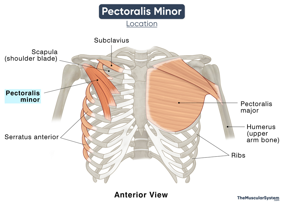
The pectoralis minor is a triangular muscle lying in the anterior thoracic region underneath its larger counterpart, the pectoralis major. It is one of the 4 anterior axioappendicular muscles, with the other 3 being the pectoralis major, serratus anterior, and subclavius. It may sometimes be referred to as ‘pecs’ minor in short.
Anatomy
Location and Attachments
| Origin | Sternal end of the 3rd, 4th, and 5th ribs, near their costal cartilages |
| Insertion | The medial border, and superior surface of the scapula’s coracoid process |
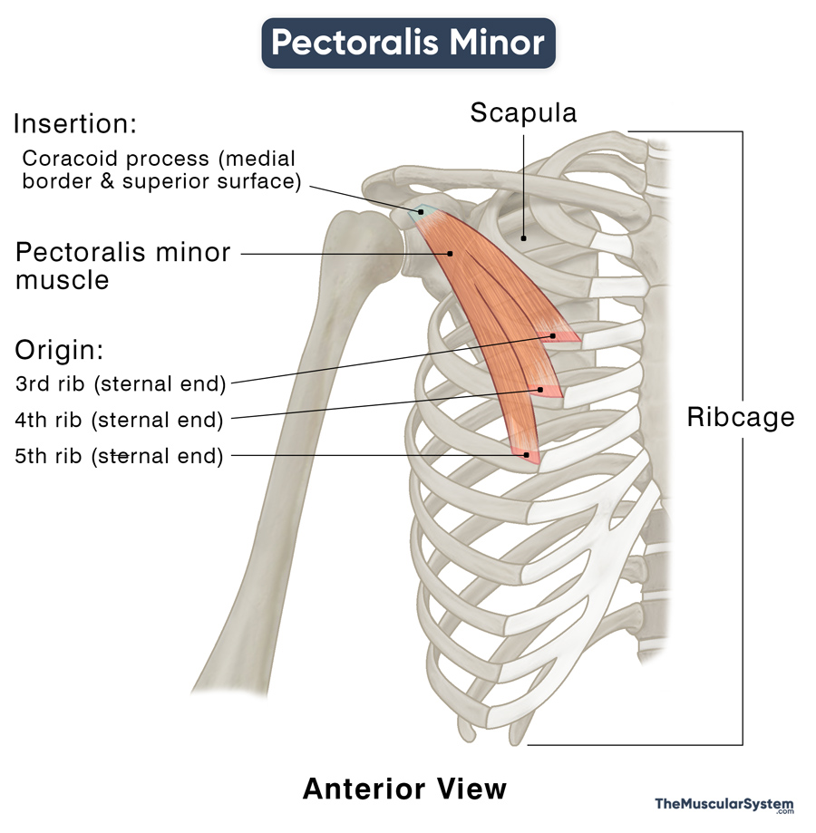
The muscle’s tendon originates as 3 heads from the 3rd to 5th ribs, lateral to their corresponding costal cartilage. The origin often extends into the fascia enclosing the adjacent intercostal muscles.
The three heads come together to form the muscle belly, coursing upward laterally, forming a flat tendon that inserts into the coracoid process.
In an anatomical variation, the muscle might originate from the 2nd rib in addition to the 3rd, 4th, and 5th.
Relations to Other Structures
The pectoralis minor is one of the most superficial muscles in the chest, with the pectoralis major being the only muscle superficial to it. The thoracoacromial artery’s pectoral branch and the lateral pectoral nerve keep these two muscles separate.
The pectoralis minor lies close to multiple fascia tissues. The clavipectoral fascia, deep to the pectoralis major, occupies the space between this muscle and the subclavius. The two pectoralis muscles, along with their associated fascia, form the front wall of the axilla.
The ribs, intercostals, and the serratus anterior are located on the posterior side of the pectoralis minor.
The muscle courses like a bridge between the rib cage and the coracoid process, letting several structures pass from the thorax to the upper arm region from underneath. The lateral, medial, and posterior cords of the brachial plexus, the axillary artery and vein are among the most important structures lying deep to the pectoralis minor. These are the primary nerve and blood supply to the forearm.
Since it overlies such vital structures, the muscle is considered an important landmark for clinical and surgical purposes. It works as a reference point for different parts of the axillary artery.
Functions
| Action | Stabilizing the scapula by pulling it down and forward against the thoracic or chest wall; elevating the 3rd to 5th ribs during inhalation |
As mentioned above, it works with the chest muscle serratus anterior to help the scapula move forward and downward against the ribcage. In this movement, the pectoralis minor acts as the synergist of the serratus anterior. This movement helps with movements at the shoulder joint, especially for extending the arm in front.
It also helps with depression of the scapula, which is usually taken care of by gravity and does not need any assistance. However, if it needs external support, the pectoralis minor is the muscle to provide it.
When the scapula is fixed, the muscle helps lift the 3rd, 4th, and 5th ribs, which in turn helps expand the ribcage. This movement allows the lungs to expand more, helping with inhalation, especially when taking a deep breath. Due to this action, the pectoralis minor can be counted as a respiratory accessory muscle.
Antagonists of the pectoralis minor are the supraspinatus, posterior deltoid, rhomboid major, teres major, levator scapulae, and upper trapezius
Innervation
| Nerve | Medial pectoral nerve (C8 and T1) |
The primary innervation comes from the medial pectoral nerve (C8, T1), a small branch of the brachial plexus (medial cord). The lateral pectoral nerve (C5, C6, C7) also innervates the muscle as a secondary source. It means the total innervation of the muscle comes from the spinal nerve roots C5 to T1.
Blood Supply
| Artery | Pectoral branch of thoracoacromial artery |
Apart from the above primary artery, the muscle also receives blood supply from the deltoid branch of the thoracoacromial artery, as well as the superior and lateral thoracic arteries.
References
- Pectoralis Minor: Function, Blood Supply & Innervation: https://study.com/academy/lesson/pectoralis-minor-function-blood-supply-innervation.html
- Pectoralis Minor: https://teachmeanatomy.info/encyclopaedia/p/pectoralis-minor/
- Pectoralis Minor Muscle: https://www.kenhub.com/en/library/anatomy/pectoralis-minor-muscle
- Pectoralis Minor Muscle – Attachment, Action & Innervation: https://www.getbodysmart.com/shoulder-muscles/pectoralis-minor-muscle/
- Pectoralis Minor Muscle: https://radiopaedia.org/articles/pectoralis-minor-muscle-1?lang=us
- Pectoralis Minor: https://rad.washington.edu/muscle-atlas/pectoralis-minor/
- Agonism and Antagonism of the Muscles of the Shoulder Joint: PracticalPainManagement.com
Della Barnes, an MS Anatomy graduate, blends medical research with accessible writing, simplifying complex anatomy for a better understanding and appreciation of human anatomy.
- Latest Posts by Della Barnes, MS Anatomy
-
Tensor Tympani
- -
Stapedius
- -
Auricularis Posterior
- All Posts


