

Levator Scapulae
By
Della Barnes, an MS Anatomy graduate, blends medical research with accessible writing, simplifying complex anatomy for a better understanding and appreciation of human anatomy.
Last updated:
02/05/2024Della Barnes, MS Anatomy
UX/UI Designer at - AdobeDella Barnes, an MS Anatomy graduate, blends medical research with accessible writing, simplifying complex anatomy for a better understanding and appreciation of human anatomy.
What is Levator Scapulae
The levator scapulae is a paired muscle in the superficial layer of extrinsic back muscles along with the trapezius, latissimus dorsi, and rhomboids.
The muscle’s name comes from its primary function of elevating the scapula, with ‘levator’ coming from the Latin word ‘levare,’ which is ‘to raise.’ Though it is anatomically a muscle of the vertebral column, functionally, it is one of the muscles controlling the scapula’s movements – the serratus posterior superior, inferior, serratus anterior, and rhomboids.
Anatomy
Location and Attachments
The paired muscle is located in the upper back, with one muscle on each side of the neck.
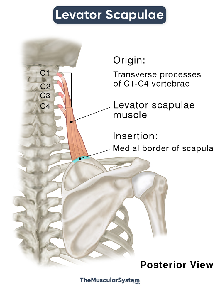
| Origin | Transverse processes of C1 to C4 vertebrae |
| Insertion | Superior side of the scapula’s medial border |
Origin
The levator scapulae originates as tendinous bands from the transverse processes of the first 4 cervical vertebrae – the atlas, axis, C3, and C4. In the case of C3 and C4, it arises from the posterior tubercles of their transverse processes.
Insertion
The muscle belly then travels downward towards the scapula to insert into the posterior lip of its medial border. The insertion point usually lies between the root of the spine of scapula and the superior scapular angle.
Relations With Surrounding Muscles and Structures
It is located superior to the major and minor rhomboids, with the sternocleidomastoid and splenius capitis muscles covering its upper part. The trapezius covers the inferior aspect of the muscle, with its middle portion being the only part to remain unobstructed. So, palpating the levator scapulae in this region is the easiest.
Additionally, the middle part of the levator scapulae contributes to the formation of the floor of the posterior neck triangle. It is an important anatomical area on the side of the neck, containing nerves and blood vessels.
Anatomical Variation
The most commonly reported anatomical variation involves changes in the points of origin and insertion. It may be attached to different numbers of vertebrae. Muscular slips may also extend to the mastoid or occipital regions or the 1st or 2nd ribs. Another variation may have it forming attachments with other muscles in the area, like the trapezius, scalene, and serratus anterior.
Function
| Action | Elevating the scapula, and rotating it so the glenoid cavity can tilt downwards |
On the Scapula and Glenoid Cavity
Elevation of the scapula is its primary function, where it works alongside the trapezius and the two rhomboids. It allows for movements like shrugging.
It is one of the primary muscles that stabilize the scapula and holds it in place during shoulder and upper arm movements.
The muscle also rotates the scapula downward, which helps tilt the glenoid cavity. During this movement, the other 4 spine muscles, the trapezius, latissimus dorsi, and rhomboids, and the thoracic wall muscles, pectoralis major and minor, also contract.
Its actions allow the shoulder girdle to retract, or draw medially back, at the scapulothoracic joint. Simultaneously, it prevents the girdle from depressing when carrying heavy objects in your hand. The levator scapulea and the rhomboids major and minor are highly active when you extend your hand toward the back, like when reaching for a back pocket.
On the Cervical Spine
The levator scapulae also acts on the cervical spine. When only the left or the right muscle contracts (unilateral contraction), it results in the tilting of the neck towards the respective side (ipsilateral rotation of the neck). You can extend the neck when both the left and right levator scapulae contract.
Antagonists
The muscle has multiple antagonists, including the shoulder muscles, anterior and posterior deltoids, teres minor and infraspinatus, the spine muscle, latissimus dorsi, and the thoracic wall muscles, pectoralis major and minor, as well as the serratus anterior.
Innervation
| Nerve | Dorsal scapular nerve (C5) and cervical nerves (C3, C4) |
Its innervation comes from the dorsal scapular nerve, arising from the anterior primary rami of the C5 nerve root from the upper part of the brachial plexus. The same nerve also innervates the two rhomboids.
The cervical nerves arising from nerve roots C3 and C4 also innervate the muscle.
Blood Supply
| Artery | Dorsal scapular artery |
Primary blood supply comes from the dorsal scapular artery.
The thyrocervical branch of the subclavian artery divides into the transverse and ascending cervical arteries. The transverse cervical artery then divides into two branches at the level of the levator scapulae to supply to it. The deep branch of the transverse cervical artery is called the dorsal scapular artery.
The vertebral artery provides additional blood supply for the vertebral part of the muscle.
References
- Anatomy, Head and Neck, Levator Scapulae Muscles: NCBI.NLM.NIH.gov
- Levator Scapulae Muscle: ScienceDirect.com
- Levator Scapulae: Osmosis.org
- Levator Scapulae Muscle: KenHub.com
- Levator Scapulae: TeachMeAnatomy.info
- Levator Scapulae: IMAIOS.com
- Agonism and Antagonism of the Muscles of the Shoulder Joint: An SEMG Approach: PracticalPainManagement.com
Della Barnes, an MS Anatomy graduate, blends medical research with accessible writing, simplifying complex anatomy for a better understanding and appreciation of human anatomy.
- Latest Posts by Della Barnes, MS Anatomy
-
Tensor Tympani
- -
Stapedius
- -
Auricularis Posterior
- All Posts


