

Serratus Posterior Superior
By
Della Barnes, an MS Anatomy graduate, blends medical research with accessible writing, simplifying complex anatomy for a better understanding and appreciation of human anatomy.
Last updated:
13/06/2024Della Barnes, MS Anatomy
UX/UI Designer at - AdobeDella Barnes, an MS Anatomy graduate, blends medical research with accessible writing, simplifying complex anatomy for a better understanding and appreciation of human anatomy.
What is the Serratus Posterior Superior
The serratus posterior superior, or SPS, is a thin rectangular extrinsic muscle located in the upper back. Together with the serratus posterior inferior, it forms the intermediate layer of back muscles. The muscle originates from the vertebral column but can still be grouped with the chest muscles because of its insertion into the ribcage. Both muscles are named after their serrated or toothed shape and assist in moving the ribcage during respiration.
Anatomy
Location and Attachments
| Origin | The nuchal ligament and the spinous process of the C7 to T3 vertebrae |
| Insertion | Superior borders of the 2nd to 5th ribs |
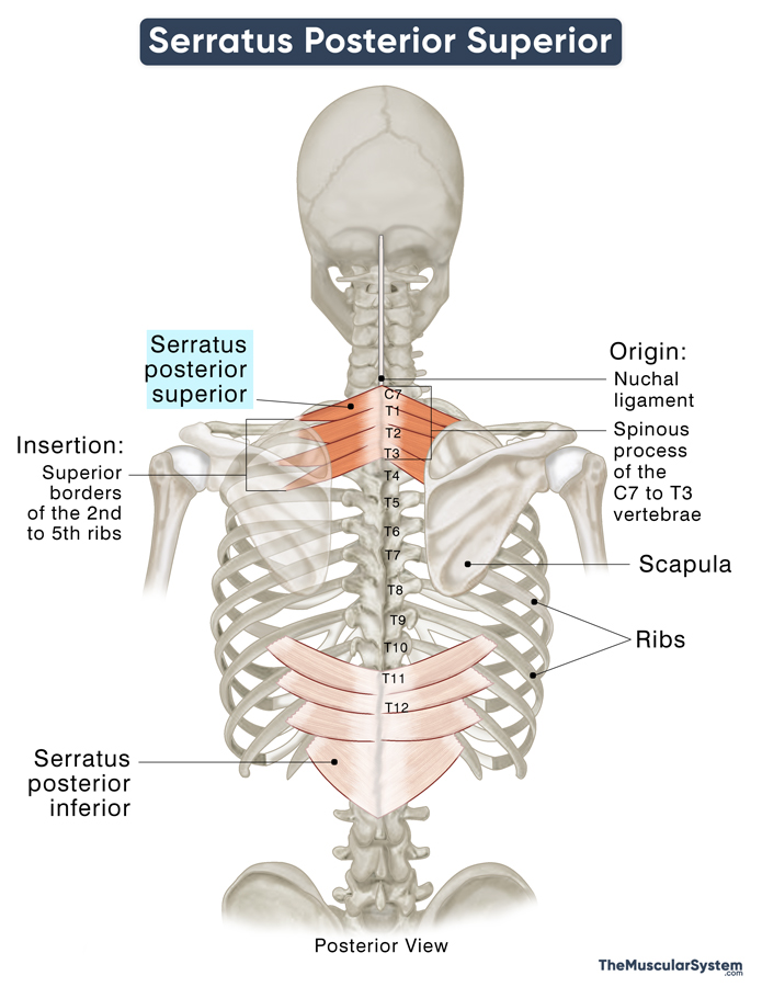
Origin
The muscle originates as a broad yet thin aponeurosis from multiple points. The lower part of the nuchal ligament and the spinous processes of the 7th cervical through the 3rd thoracic vertebrae are the primary points of origin. Some part of the muscle also arises from the adjacent supraspinous ligaments.
Insertion
The tendinous sheath becomes more muscular as it courses laterally downward the back of the thoracic cage. The muscle then forms 4 fleshy finger-like projections that insert into the superior borders and outer surfaces of the 2nd to 5th ribs, just lateral to their costal angle.
These finger-like projections at its insertion point make the muscle look ‘toothed’ or serrated.
Anatomical Variation
The muscle may show some variation in these origin and insertion points. It may originate from only the C7 to up to the T2 vertebra and insert into the 2nd-4th ribs instead of the 2nd-5th.
Relations With Surrounding Muscles and Structures
The rhomboids major and minor, and the trapezius muscle lie superficial to the serratus posterior superior, while the two splenius muscles and the erector spinae muscles lie deep to it.
The muscle is also superficial to the thoracolumbar fascia in the thoracic region. On its lateral side lies the levator scapulae muscle.
Function
| Action | Elevating the superior ribs to aid in inhalation |
Though there is some debate over the actual function of the serratus posterior, this muscle is believed to help elevate the superior portion of the ribcage by raising the ribs to which it is attached. This action supports respiration, particularly during inhalation, as it expands the thoracic cavity, allowing air to enter the lungs.
Innervation
| Nerve | 2nd to 5th Intercostal nerves (T2-T5) |
Its innervation comes from the intercostal nerves T2 to T5. These are the ventral or anterior rami of the 2nd to 5th thoracic spinal nerves.
Blood Supply
| Artery | Posterior intercostal arteries |
The muscle gets its blood supply from the dorsal branches of the posterior intercostal arteries.
References
- Serratus Posterior Superior: TeachMeAnatomy.info
- Serratus Posterior Superior Muscle: Elsevier.com
- Serratus Posterior Superior Muscle: IMAIOS.com
- Serratus Posterior Superior Muscle: RadioPaedia.org
- Serratus Posterior Muscles: Study.com
- Serratus Posterior Muscles: Anatomy, Clinical Relevance, And Function: PubMed.NCBI.NLM.NIH.gov
- Serratus Posterior Muscles: Kenhub.com
Della Barnes, an MS Anatomy graduate, blends medical research with accessible writing, simplifying complex anatomy for a better understanding and appreciation of human anatomy.
- Latest Posts by Della Barnes, MS Anatomy
-
Tensor Tympani
- -
Stapedius
- -
Auricularis Posterior
- All Posts


