

Erector Spinae
By
Della Barnes, an MS Anatomy graduate, blends medical research with accessible writing, simplifying complex anatomy for a better understanding and appreciation of human anatomy.
Last updated:
01/10/2024Della Barnes, an MS Anatomy graduate, blends medical research with accessible writing, simplifying complex anatomy for a better understanding and appreciation of human anatomy.
What Is the Erector Spinae Group
The erector spinae, also known as the sacrospinalis, is a group of muscles comprising the middle or intermediate layer of the intrinsic or deep back muscles. The name literally means ‘spinal erectors,’ with the group including 3 muscles that span the entire length of the spinal column.
The thin, long bands of muscles are the primary extensors of the back and are instrumental in the movements and flexibility of the back and neck.
Names and Basic Anatomy of the Erector Spinae Muscles
The erector spinae muscle group is located along the lateral side of the back, between the spinous processes of the vertebrae and the ribs’ costal angles. It consists of 3 primary muscles, each divided into 3 parts, resulting in a total of 9 muscles in the group.
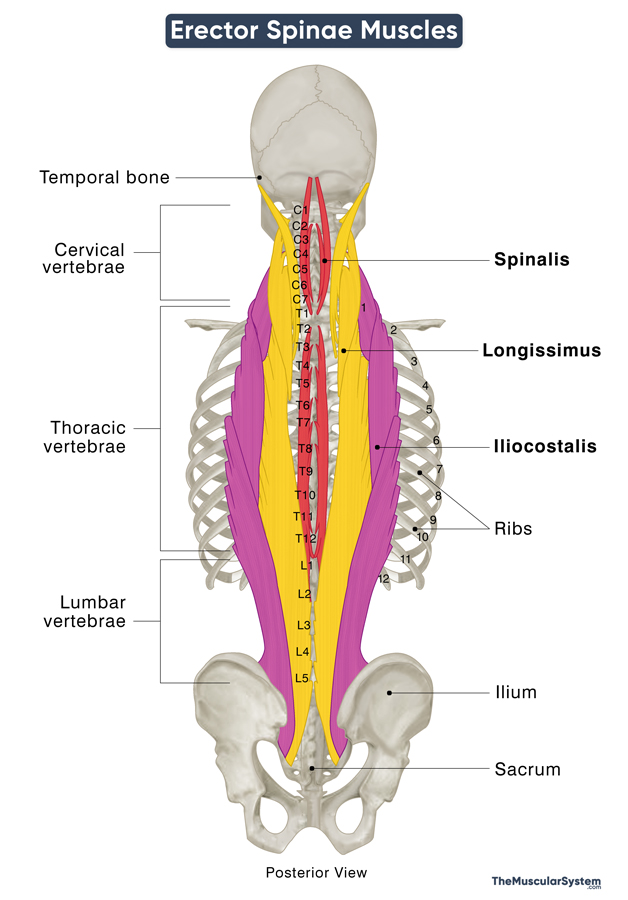
The 3 main erector spinae muscles, listed from deepest to most superficial, are as follows:
| Name | Divisions | Origin | Insertion | Actions | Innervation | Blood Supply |
|---|---|---|---|---|---|---|
| 1. Spinalis The smallest and most medially located of the three | – Spinalis Capitis (at the base of the skull) – Spinalis Cervicis (at the back of the neck) – Spinalis Thoracis (in the upper to mid back) | Spinous processes of the 7th cervical to 2nd lumbar vertebrae (C7-L2), and the nuchal ligament | Spinous processes of the 2nd cervical to the 8th thoracic vertebrae (C2-T8); some part of the muscle blends with the semispinalis capitis muscle | – Unilateral contractions flex the spinal cord sideways, helping bend the body left or right – Bilateral contractions straighten the back and neck and also bend it backward | Dorsal rami of the cervical, thoracic, and lumbar spinal nerves corresponding to the levels of each part of the muscle | Deep cervical and vertebral arteries; dorsal branches of supreme and posterior intercostal arteries |
| 2. Longissimus The largest of the three muscles, located between the other two | – Longissimus Capitis (at the base of the skull) – Longissimus Cervicis (at lower neck and upper back) – Longissimus Thoracis (in the mid to lower back) | Transverse processes of the 4th cervical to 5th lumbar vertebrae (C4-L5), and the ilium and sacrum bones | Mastoid process of the temporal bone and transverse processes of 2nd cervical to 5th lumbar vertebrae (C2-L5) | Both unilateral and bilateral contractions contribute to the same movements as above – but being the largest of the 3 muscles in the group, it is the primary contributor to these movements. | Dorsal rami of the corresponding branches of cervical, thoracic, and lumbar spinal nerves | Vertebral, occipital, and cervical arteries; sacral, and intercostal arteries |
| 3. Iliocostalis The most laterally located of the three | – Iliocostalis Cervicis (at the back of the neck) – Iliocostalis Thoracis (in the upper to mid back) – Iliocostalis Lumborum (in the mid to lower back) | Angles of the 3rd to 12th ribs, the lateral sacral, and iliac crests | Transverse processes of the 4th cervical to 4th lumbar vertebrae and the costal angles of all the ribs | Same as the other two erector spinae muscles | Dorsal rami of the corresponding branches of cervical, thoracic, and lumbar spinal nerves | Deep cervical, occipital, and vertebral arteries; posterior intercostal and subcostal arteries; Lateral sacral and lumbar arteries |
Relations With Surrounding Muscles and Structures
The erector spinae muscles are deep to the serratus posterior inferior, the two rhomboids, and the splenius capitis and cervicis muscles. They also have the thoracolumbar fascia lying superficially.
The tendons of the thoracic fibers of the longissimus and iliocostalis lumborum, along with the superficial fibers of the multifidus muscle, converge to form the erector spinae aponeurosis.
Antagonists
The rectus abdominis is an abdominal muscle that extends from the ribs to the pubic bones at the front of the body. Since it helps bend the body forward, it is antagonistic to the erector spinae muscles as they help straighten the back and bend it backward.
References
- Anatomy, Back, Muscles: NCBI.NLM.NIH.gov
- Erector Spinae Muscles: Kenhub.com
- The Intrinsic Back Muscles: TeachMeAnatomy.info
- Erector Spinae Group: Radiopaedia.org
- Erector Spinae Muscle | Pain, Action & Origin: Study.com
Della Barnes, an MS Anatomy graduate, blends medical research with accessible writing, simplifying complex anatomy for a better understanding and appreciation of human anatomy.
- Latest Posts by staff All Posts


