

Spinalis
By
Della Barnes, an MS Anatomy graduate, blends medical research with accessible writing, simplifying complex anatomy for a better understanding and appreciation of human anatomy.
Last updated:
14/10/2024Della Barnes, MS Anatomy
UX/UI Designer at - AdobeDella Barnes, an MS Anatomy graduate, blends medical research with accessible writing, simplifying complex anatomy for a better understanding and appreciation of human anatomy.
What Is the Spinalis
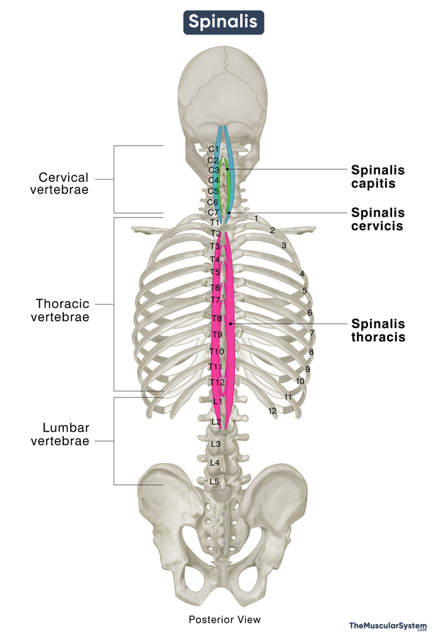
The spinalis is the most medially located muscle in the erector spinae group, which forms the intermediate layer of the deep intrinsic back muscles. The other two muscles in this group are the longissimus and Iliocostalis.
The spinalis is positioned closest to the spinal cord out of all three erector spinae muscles and plays a vital role in the movements of the vertebral column.
Anatomy
Location and Attachments
It consists of a bundle of thin, long muscle fibers or fascicles that vary in length and their points of attachment. The muscle can be divided into three parts, with each part having different points of origin and insertion:
| Origin | Spinalis Capitis: Spinous processes of C7 and T1 vertebrae via the inserting fibers of the semispinalis capitis muscle Spinalis Cervicis: Spinous process of C7 vertebrae and the nuchal ligament Spinalis Thoracis: Spinous processes of T11 to L2 vertebrae |
| Insertion | Spinalis Capitis: No clear insertion point as it blends with the semispinalis capitis muscle Spinalis Cervicis: Spinous processes of C2 to C4 vertebrae Spinalis Thoracis: Spinous processes of the upper thoracic vertebrae (usually down to T8) |
The muscle bundles that course medially have lower origins and higher insertions, making them shorter, while the lateral bundles have higher origins and lower insertions, making them longer.
Spinalis Capitis
The least prominent yet topmost part of the spinalis muscle, the spinalis capitis is no more than an extension of the semispinalis capitis (another deep back muscle), originating from the 7th cervical to the 1st thoracic vertebrae (C7 to T1). The name ‘capitis’ comes from the fact that this part of the spinalis attaches to the base of the skull via the semispinalis capitis, which inserts into the occipital bone.
Spinalis Cervicis
The spinalis cervicis is formed of poorly developed, shorter muscle fibers. They originate from the spinous process of the 6th or 7th cervical vertebrae, with some fibers also arising from the caudal part of the adjacent nuchal ligament. From their point of origin, the fibers travel upward toward the cranium to insert into the spinous processes of the Axis (C2) and the 3rd and 4th cervical vertebrae (C3 and C4).
Spinalis Thoracis
It is the most prominent part of the spinalis muscle, with its long fascicles running laterally. They originate from the spinous processes of the 11th thoracic vertebrae (T11) to the 2nd lumbar vertebrae (L2). Then the fibers travel upward all the way to insert into the spinous processes of the 2nd or 3rd to the 8th thoracic vertebrae (T2 or T3 to T8)
Relations With Surrounding Muscles and Structures
The thoracic part of the muscle is located on the medial side of the longissimus muscle, and the two often blend completely. Since the spinalis is a muscle in the deep layer, the thoracic spinalis lies deep to the latissimus dorsi, serratus posterior inferior, and the lower part of the trapezius.
The point of insertion of the spinalis cervicis on the 2nd cervical vertebrae lies inferior to the insertion points of the two neck muscles, obliquus capitis inferior and rectus capitis posterior major, which also inserts into the C2. The cervical part also has the semispinalis capitis lying lateral to it.
Function
| Action | Unilateral: Flexing the spinal column to bend the body on one side Bilateral: Extending the spine to straighten the neck and trunk |
The spinalis works with the rest of the erector spinae muscles to move and stabilize the vertebral column and pelvis. When the erector spinae muscles contract on one side (unilaterally), they cause the spine to flex laterally towards the same side (ipsilateral flexion). This action affects the entire spinal column, including the cervical, thoracic, and lumbar regions, leading to a sideward bending motion of the body.
When these muscles contract on both sides (bilaterally), the head, neck, and entire spinal column are extended. It means the spine bends backward or straightens from a flexed position.
Innervation
| Nerve | Posterior or dorsal rami of the spinal nerves |
Each part of the spinalis muscle gets its innervation from the lateral branches of the posterior rami of the corresponding cervical, thoracic, and lumbar spinal nerves.
Blood Supply
| Artery | The deep cervical and vertebral arteries, and the dorsal branches of the supreme and posterior intercostal arteries |
The different parts of the muscle receive blood supply from different sources.
The cervicis and capitis parts are supplied by the muscular branches of the vertebral and deep cervical arteries. The descending branches of the occipital artery also contribute to the vasculature in this part.
The thoracis part gets its blood supply from the dorsal branches of the supreme and posterior intercostal arteries. The lower part of the spinalis thoracis that attaches to the lumbar vertebra gets blood supply from the lumbar arterial branches.
References
- Spinalis: GPNotebook.com
- Erector Spinae Muscle | Pain, Action & Origin: Study.com
- Spinalis Muscle: Kenhub.com
- Spinalis: TeachMeAnatomy.info
- Spinalis Thoracis Muscle: Elsevier.com
- Anatomy, Back, Muscles: NCBI.NLM.NIH.gov
Della Barnes, an MS Anatomy graduate, blends medical research with accessible writing, simplifying complex anatomy for a better understanding and appreciation of human anatomy.
- Latest Posts by Della Barnes, MS Anatomy
-
Tensor Tympani
- -
Stapedius
- -
Auricularis Posterior
- All Posts


