

Iliacus
By
Della Barnes, an MS Anatomy graduate, blends medical research with accessible writing, simplifying complex anatomy for a better understanding and appreciation of human anatomy.
Last updated:
14/05/2025Della Barnes, MS Anatomy
UX/UI Designer at - AdobeDella Barnes, an MS Anatomy graduate, blends medical research with accessible writing, simplifying complex anatomy for a better understanding and appreciation of human anatomy.
What is the Iliacus
The Iliacus is a flat, fan-shaped, paired muscle in the group of posterior abdominal muscles. Located in the iliac fossa, it is part of the iliopsoas muscle complex, which also includes the psoas major.
The muscle is anatomically grouped with the posterior abdominal wall muscles, although it is often functionally and spatially associated with the pelvis and hip due to its significant role in hip stability and movement.
Anatomy
Location and Attachments
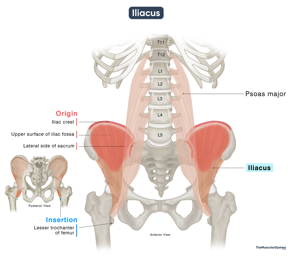
| Origin | Upper surface of the iliac fossa, the iliac crest, lateral side of the sacrum bone |
| Insertion | Lesser trochanter on the proximal femur (via iliopsoas) |
Origin
The iliacus muscle originates from a broad region within the pelvis, with its primary attachment being the upper two-thirds of the iliac fossa, which is the wide, concave inner surface of the ilium’s wing. This broad attachment gives the muscle a strong and stable foundation across the pelvic region.
In addition to the iliac fossa, portions of the muscle also arise from the inner lip of the iliac crest, particularly along its anterior border. The muscle further extends its attachment to the anterolateral surface of the sacrum, adding to its structural base.
Apart from these bony attachments, the iliacus also draws fibers from nearby ligaments, including the anterior sacroiliac and iliolumbar ligaments.
Insertion
From its origin, the muscle courses downward and forward to join the fibers of the psoas major near the level of the hip joint capsule. This is where the iliopsoas muscle forms.
As the combined fibers continue to course downward, they become tendinous and pass under the inguinal ligament through a small opening (the muscular lacuna) to reach the front of the thigh. Here, they insert onto the lesser trochanter of the femur (thigh bone). Within this common tendon, the iliacus fibers are positioned slightly anterior to those of the psoas major.
Relations With Surrounding Muscles and Structures
In the Pelvic Region
Within the pelvis, the iliacus is enveloped by the iliac fascia, a layer of connective tissue that separates it from the abdominal peritoneum. The lateral femoral cutaneous nerve (L2-L3), which supplies sensation to the anterolateral thigh, courses obliquely across the anterior surface of the iliacus
On the right side, the cecum, which is the beginning portion of the large intestine, lies anterior to the right iliacus muscle.
On the left side, the iliac colon (the part of the descending colon located in the left iliac fossa) lies anterior to the left iliacus muscle.
The psoas major muscle lies medial to the iliacus before the two muscles merge.
In the Thigh Region
It sits behind the thigh muscles, rectus femoris and sartorius, and the fascia lata — the thick sheet of connective tissue that covers the musculature of the thigh.
The hip joint lies behind the iliacus muscle, with the large iliopsoas bursa separating the two. The deep femoral artery, one of the main arteries supplying the thigh and lower limb, crosses over the muscle in this region.
Function
| Action | Flexing the thigh and torso at the hip |
The muscle works with the psoas major, and as part of the iliopsoas, it is responsible for some of the major movements of the hips and thigh.
When the pelvis and torso are held still, the muscle’s contraction helps flex the thigh, bringing it toward the body. An example is when you bring your knees closer to the torso while sitting in a chair. The muscle also contributes to the lateral rotation of the thigh at the hip joint.
On the other hand, when the thigh is fixed, the muscle contracts and helps with flexing the trunk. An example would be when you bend forward or do a sit-up.
These movements are important for activities like walking, running, and jumping. Additionally, the muscle contributes to the stability of the pelvis and hip.
Antagonists
The gluteus maximus, the main muscle responsible for extending the hip, works in opposition to the liacus, making them antagonistic.
Innervation
| Nerve | Femoral nerve |
The muscle receives innervation from the femoral nerve, the largest division of the lumbar plexus, branching from the posterior branches of the ventral rami of the L2, L3, and L4 spinal nerves.
Blood Supply
| Artery | Lumbar branch of the iliolumbar artery |
Like the psoas major, this muscle also receives blood supply mainly from the iliolumbar artery, a branch of the posterior trunk of the internal iliac artery.
Additional blood supply comes from the obturator, deep circumflex iliac, and femoral arteries.
References:
- Iliacus Muscle: Radiopaedia.org
- Iliacus: Rad.Washington.edu
- Iliacus Muscle: Elsevier.com
- Iliacus Muscle: Kenhub.com
- Iliacus: Teachmeanatomy.info
- Iliacus Muscle: ScienceDirect.com
- Iliacus Muscle Function & Action: Study.com
Della Barnes, an MS Anatomy graduate, blends medical research with accessible writing, simplifying complex anatomy for a better understanding and appreciation of human anatomy.
- Latest Posts by Della Barnes, MS Anatomy
-
Tensor Tympani
- -
Stapedius
- -
Auricularis Posterior
- All Posts


